The study presents a volumetric electrocardiographic imaging (ECGI) methodology that estimates cardiac sources using Poisson's equation and Green's functions to relate body surface potentials to cardiac sources, enabling three-dimensional non-invasive cardiac mapping.
Volumetric Non-Invasive Cardiac Mapping for Accessible Global Arrhythmia Characterization
Publication
Metrics
Quick Answers
What methodology did the authors use?
The study presents a volumetric electrocardiographic imaging (ECGI) methodology that estimates cardiac sources using Poisson's equation and Green's functions to relate body surface potentials to cardiac sources, enabling three-dimensional non-invasive cardiac mapping. More in Methodology →
What are the key results?
The method achieved accurate activation time mapping in both simulated and clinical scenarios — It demonstrated improved localization accuracy in complex anatomical regions like the septum and ventricular base More in Key Results →
Why is this work significant?
This research advances non-invasive cardiac electrophysiology by providing a robust framework for accurate activation mapping, which could enhance diagnostic precision and treatment planning for arrhythmias. More in Significance →
What are the main limitations?
The method relies on simplified conductivity assumptions which may introduce systematic errors — It requires further validation in diverse patient populations with varying cardiac geometries More in Limitations →
Abstract
Cardiac arrhythmias are a major cause of morbidity and mortality increasing the risk of stroke, heart failure, and sudden cardiac death. Imageless electrocardiographic imaging (ECGI) provides a non invasive alternative to electrical mapping from body surface potentials, but conventional ECGI is confined to epicardial reconstructions and can miss arrhythmias originating in deeper myocardium. We address this by reconstructing three dimensional cardiac activity with a volumetric formulation that solves an inverse source problem via Green's functions, enabling full volume activation mapping and improved localization in anatomically complex regions. We evaluate the approach on simulated premature ventricular beats and on four challenging patient cases, a right ventricular outflow tract premature ventricular contraction, a left bundle branch block, a ventricular tachycardia, and Wolff Parkinson White, and additionally assess performance on an open source myocardial infarction dataset. Results show that volumetric ECGI recovers 3D activation and sharpens arrhythmia origin localization, achieving a 59.3% reduction in geodesic error between estimated and simulated origins relative to surface only methods; in patient cases, activation patterns align with clinical diagnoses. Overall, imageless volumetric ECGI offers accessible, non invasive 3D activation mapping that overcomes a core limitation of surface restricted techniques and may improve preprocedural planning, ablation target guidance, and selection or optimization of cardiac resynchronization therapy.
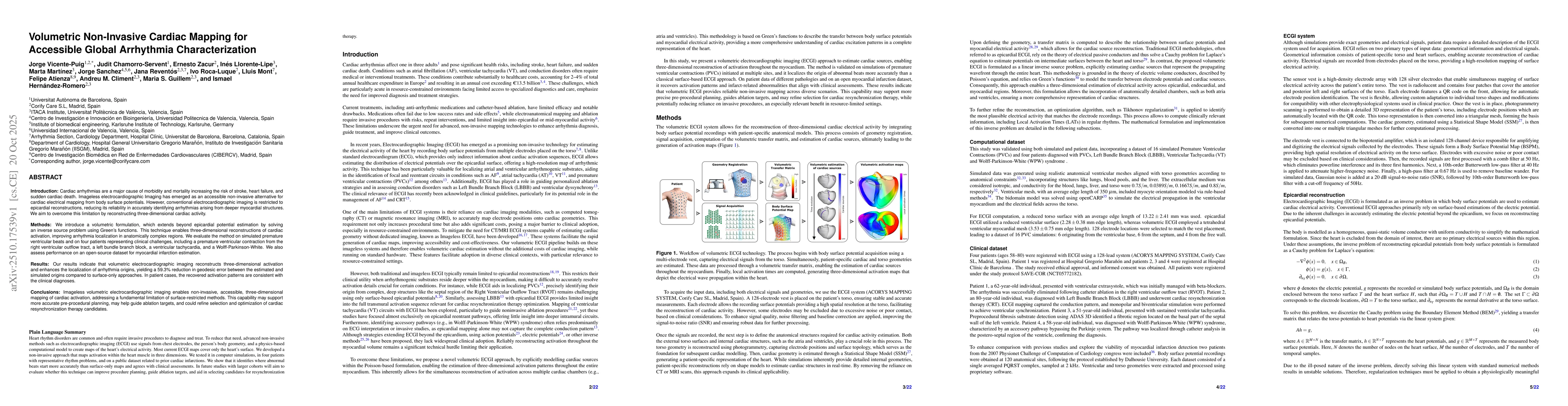
Key Findings, in focus
Seven facets of this paper, analysed and brought into focus by AI.
This research advances non-invasive cardiac electrophysiology by providing a robust framework for accurate activation mapping, which could enhance diagnostic precision and treatment planning for arrhythmias.
- The method achieved accurate activation time mapping in both simulated and clinical scenarios
- It demonstrated improved localization accuracy in complex anatomical regions like the septum and ventricular base
- The approach showed potential for guiding ablation therapies and improving CRT patient selection
This research advances non-invasive cardiac electrophysiology by providing a robust framework for accurate activation mapping, which could enhance diagnostic precision and treatment planning for arrhythmias.
Development of a volumetric source imaging technique using Green's functions for direct estimation of activation times from body surface potentials
The approach explicitly solves Poisson's equation for cardiac source estimation and uses Green's functions to establish a physiologically meaningful relationship between body surface potentials and cardiac sources, differing from traditional epicardial-focused ECGI methods
- The method relies on simplified conductivity assumptions which may introduce systematic errors
- It requires further validation in diverse patient populations with varying cardiac geometries
- Incorporate anatomical priors and spatially varying conductivity models to improve accuracy
- Conduct prospective trials with invasive ground truth to quantify performance
Discussion 0