Authors
Summary
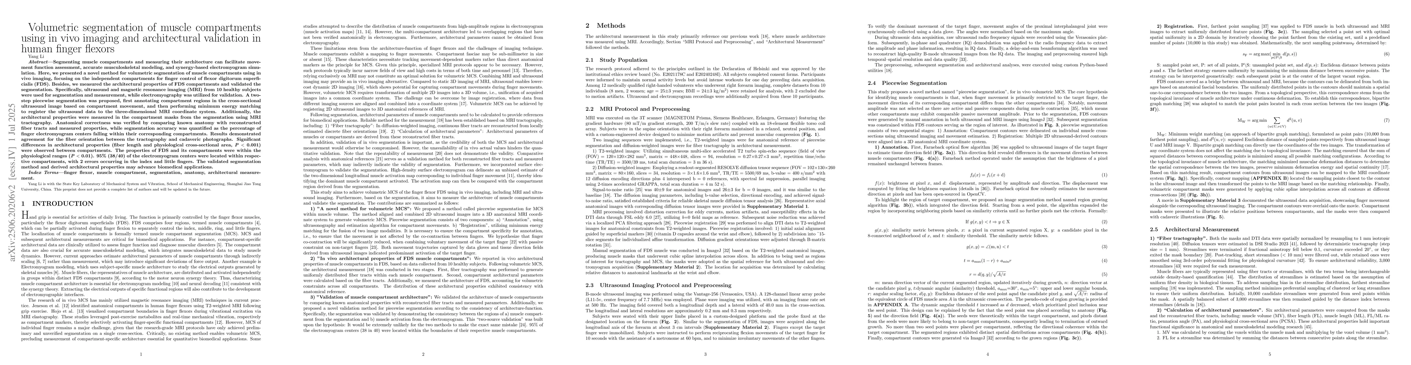
Segmenting muscle compartments and measuring their architecture can facilitate movement function assessment, accurate musculoskeletal modeling, and synergy-based electromyogram simulation. Here, we presented a novel method for volumetric segmentation of muscle compartments using in vivo imaging, focusing on the independent compartments for finger control of flexor digitorum superficialis (FDS). Besides, we measured the architectural properties of FDS compartments and validated the segmentation. Specifically, ultrasound and magnetic resonance imaging (MRI) from 10 healthy subjects were used for segmentation and measurement, while electromyography was utilized for validation. A two-step piecewise segmentation was proposed, first annotating compartment regions in the cross-sectional ultrasound image based on compartment movement, and then performing minimum energy matching to register the ultrasound data to the three-dimensional MRI coordinate system. Additionally, the architectural properties were measured in the compartment masks from the segmentation using MRI tractography. Anatomical correctness was verified by comparing known anatomy with reconstructed fiber tracts and measured properties, while segmentation accuracy was quantified as the percentage of finger electromyogram centers falling within their corresponding compartments. Results demonstrated agreement for the fiber orientation between the tractography and cadaveric photographs. Significant differences in architectural properties (P < 0.001) were observed between compartments. The properties of FDS and its compartments were within the physiological ranges (P < 0.01). 95% (38/40) of the electromyogram centers were located within respective compartments, with 2 errors occurring in the index and little fingers. The validated segmentation method and derived architectural properties may advance biomedical applications.
AI Key Findings
Get AI-generated insights about this paper's methodology, results, and significance.
Paper Details
PDF Preview
Citation Network
Current paper (gray), citations (green), references (blue)
Display is limited for performance on very large graphs.
Similar Papers
Found 4 papersNo citations found for this paper.
Comments (0)