Summary

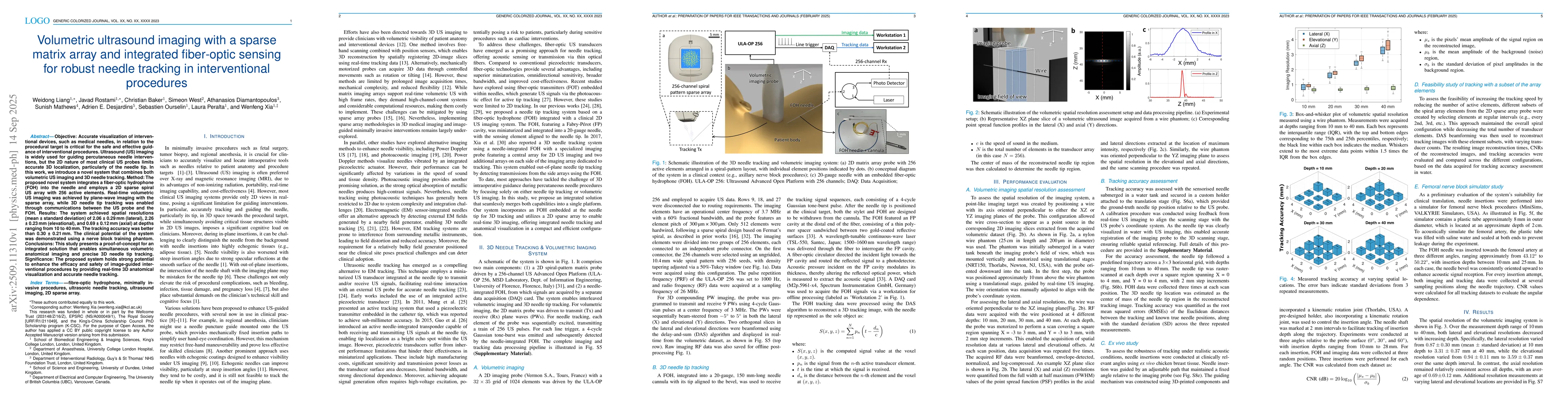
Accurate visualization of interventional devices, such as medical needles, is essential for the safe and effective guidance of minimally invasive procedures. Ultrasound (US) imaging is widely used for needle guidance, but the two-dimensional nature of most clinical probes limits accurate three-dimensional (3D) localization, particularly of the needle tip. We present a novel system that integrates volumetric US imaging with 3D needle tracking by combining a fiber-optic hydrophone embedded in the needle and a sparse spiral US array. Real-time volumetric imaging was achieved using plane-wave techniques, while precise needle tip tracking was enabled through communication between the probe and hydrophone. The feasibility of the approach was demonstrated using a nerve block training phantom. This proof-of-concept system enables simultaneous volumetric anatomical imaging and 3D needle tip tracking, with strong potential to enhance the efficacy and safety of image-guided interventional procedures.

AI Key Findings

Generated Oct 02, 2025

Methodology

The study combines volumetric ultrasound imaging with a sparse spiral matrix array and fiber-optic hydrophone (FOH) integrated into the needle for real-time 3D needle tip tracking. The system uses plane-wave imaging for volumetric data acquisition and FOH for precise tip localization, validated using phantoms and ex vivo tissue.

Key Results

  • The system achieved sub-millimeter tracking accuracy with mean errors of 0.11-0.20 mm at depths of 10-4,0 mm.
  • Tracking accuracy decreased with increasing distance from the probe center and imaging depth, showing a mean error of 1.30 mm at the periphery.
  • CNR of tracking images dropped from 29.99 dB at 0° insertion angle to 9.26 dB at 60°, due to reduced FOH sensitivity.

Significance

This integration enables simultaneous 3D anatomical imaging and accurate needle tip tracking, enhancing safety and precision in image-guided interventional procedures, particularly when the needle is not visible in standard ultrasound.

Technical Contribution

Development of a 2D sparse spiral matrix array combined with fiber-optic sensing for real-time volumetric imaging and 3D needle tracking, offering improved spatial resolution and robust tracking performance.

Novelty

First integrated system combining volumetric US imaging with 3D needle tracking using a sparse matrix array and fiber-optic hydrophone, enabling simultaneous anatomical visualization and precise needle localization.

Limitations

  • Tracking accuracy degrades significantly with reduced number of active elements (e.g., 30 elements led to 8.11 mm error).
  • Processing time for 3D tracking images remains high (22.79 seconds per frame), limiting real-time intraoperative use.

Future Work

  • Optimize computational efficiency through GPU acceleration and reduced reconstruction volumes.
  • Explore coherent multi-transducer ultrasound imaging to improve spatial resolution and field-of-view without sacrificing frame rate.

Paper Details

PDF Preview

Download PDF

Comments (0)

This website uses cookies to ensure you get the best experience. Learn more