Vortex Interference Enables optimal 3D Interferometric Nanoscopy
Publication
Metrics
AI Quick Summary
Vortex Interference Localization Microscopy (VILM) simplifies super-resolution imaging by using a pair of vortex phase plates to encode axial position via bilobed detection PSFs, achieving optimal 3D nanoscopy with a single output image, thus reducing instrumentation complexity. This streamlined approach maintains ultra-high 3D precision and resolves microtubule architecture and signaling proteins in cell adhesions.
Paper Preview
Abstract
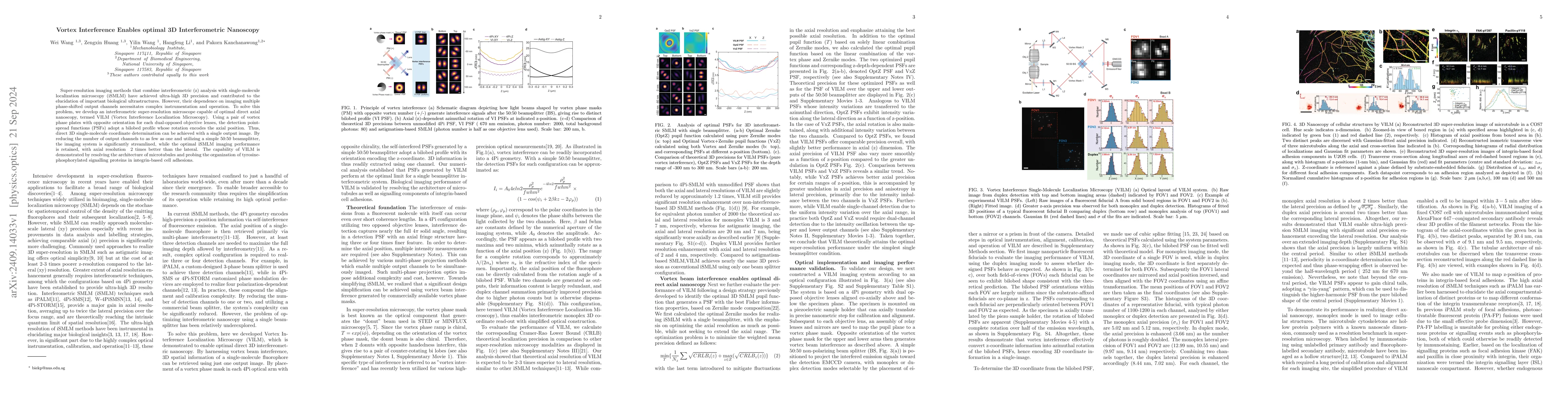
Super-resolution imaging methods that combine interferometric (z) analysis with single-molecule localization microscopy (iSMLM) have achieved ultra-high 3D precision and contributed to the elucidation of important biological ultrastructures. However, their dependence on imaging multiple phase-shifted output channels necessitates complex instrumentation and operation. To solve this problem, we develop an interferometric super-resolution microscope capable of optimal direct axial nanoscopy, termed VILM (Vortex Interference Localization Microscopy). Using a pair of vortex phase plates with opposite orientation for each dual-opposed objective lenses, the detection point-spread functions (PSFs) adopt a bilobed profile whose rotation encodes the axial position. Thus, direct 3D single-molecule coordinate determination can be achieved with a single output image. By reducing the number of output channels to as few as one and utilizing a simple 50:50 beamsplitter, the imaging system is significantly streamlined, while the optimal iSMLM imaging performance is retained, with axial resolution ~2 times better than the lateral. The capability of VILM is demonstrated by resolving the architecture of microtubules and probing the organization of tyrosine-phosphorylated signalling proteins in integrin-based cell adhesions.
AI Key Findings
Get AI-generated insights about this paper's methodology, results, significance, and more — seven facets brought into focus.
Impact
Authors
PDF Preview
Citation Network
Current paper (gray), citations (green), references (blue)
Display is limited for performance on very large graphs.
Discussion 0