Summary
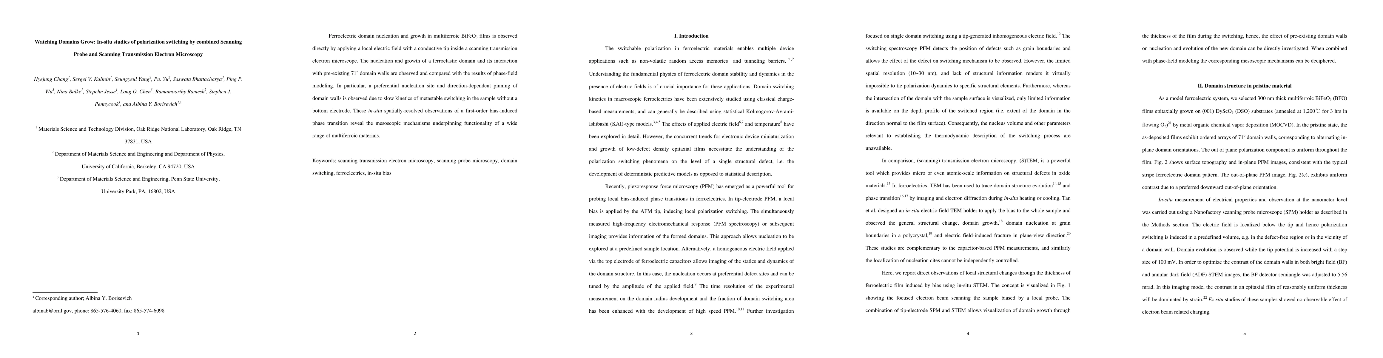
Ferroelectric domain nucleation and growth in multiferroic BiFeO3 films is observed directly by applying a local electric field with a conductive tip inside a scanning transmission electron microscope. The nucleation and growth of a ferroelastic domain and its interaction with pre-existing 71^{\circ} domain walls are observed and compared with the results of phase-field modeling. In particular, a preferential nucleation site and direction-dependent pinning of domain walls is observed due to slow kinetics of metastable switching in the sample without a bottom electrode. These in-situ spatially-resolved observations of a first-order bias-induced phase transition reveal the mesoscopic mechanisms underpinning functionality of a wide range of multiferroic materials.
AI Key Findings
Get AI-generated insights about this paper's methodology, results, and significance.
Paper Details
PDF Preview
Key Terms
Citation Network
Current paper (gray), citations (green), references (blue)
Display is limited for performance on very large graphs.
Similar Papers
Found 4 papersWatching the Watchers: A Comparative Fairness Audit of Cloud-based Content Moderation Services
David Hartmann, Amin Oueslati, Dimitri Staufer
| Title | Authors | Year | Actions |
|---|
Comments (0)