WaveFlow - Towards Integration of Ultrasound Processing with Deep Learning
Publication
Metrics
AI Quick Summary
WaveFlow integrates ultrasound data acquisition and processing tools with TensorFlow for real-time ultrasound image reconstruction, achieving frame rates of 55 fps for B-mode and 17 fps for synthetic and plane-wave imaging algorithms. The framework includes operators for beamforming and signal processing optimized for both CPU and GPU.
Paper Preview
Abstract
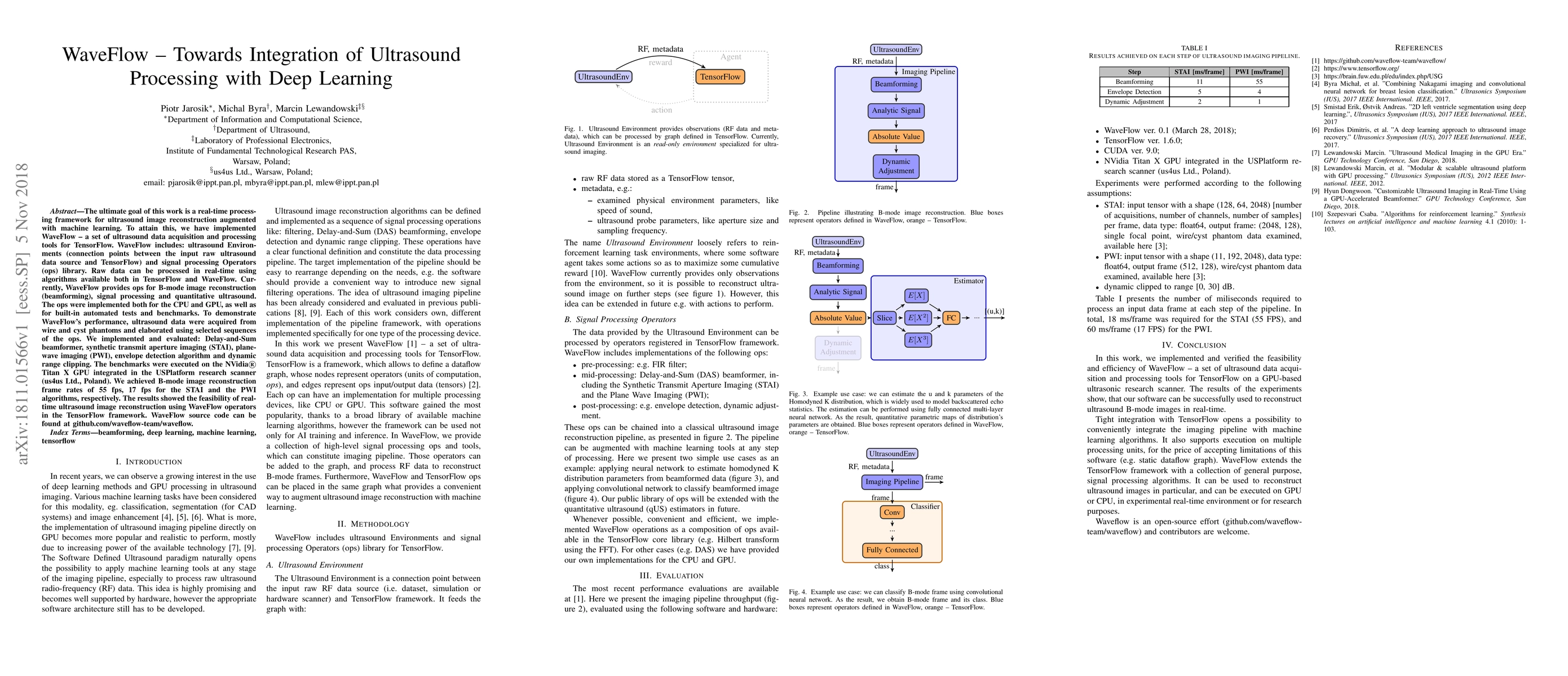
The ultimate goal of this work is a real-time processing framework for ultrasound image reconstruction augmented with machine learning. To attain this, we have implemented WaveFlow - a set of ultrasound data acquisition and processing tools for TensorFlow. WaveFlow includes: ultrasound Environments (connection points between the input raw ultrasound data source and TensorFlow) and signal processing Operators (ops) library. Raw data can be processed in real-time using algorithms available both in TensorFlow and WaveFlow. Currently, WaveFlow provides ops for B-mode image reconstruction (beamforming), signal processing and quantitative ultrasound. The ops were implemented both for the CPU and GPU, as well as for built-in automated tests and benchmarks. To demonstrate WaveFlow's performance, ultrasound data were acquired from wire and cyst phantoms and elaborated using selected sequences of the ops. We implemented and evaluated: Delay-and-Sum beamformer, synthetic transmit aperture imaging (STAI), plane-wave imaging (PWI), envelope detection algorithm and dynamic range clipping. The benchmarks were executed on the NVidia Titan X GPU integrated in the USPlatform research scanner (us4us Ltd., Poland). We achieved B-mode image reconstruction frame rates of 55 fps, 17 fps for the STAI and the PWI algorithms, respectively. The results showed the feasibility of real-time ultrasound image reconstruction using WaveFlow operators in the TensorFlow framework. WaveFlow source code can be found at github.com/waveflow-team/waveflow
AI Key Findings
Get AI-generated insights about this paper's methodology, results, significance, and more — seven facets brought into focus.
Impact
Paper Details
PDF Preview
Key Terms
Citation Network
Current paper (gray), citations (green), references (blue)
Display is limited for performance on very large graphs.
Discussion 0