Wavelet leader based formalism to compute multifractal features for classifying lung nodules in X-ray images
Publication
Metrics
AI Quick Summary
This paper introduces a novel lung nodule classification algorithm using multifractal features derived from X-ray images, employing wavelet leader formalism and a Support Vector Machine classifier. The method combines wavelet-based pre-processing and classical texture features for enhanced accuracy, achieving a maximum ROC AUC of 75%.
Paper Preview
Abstract
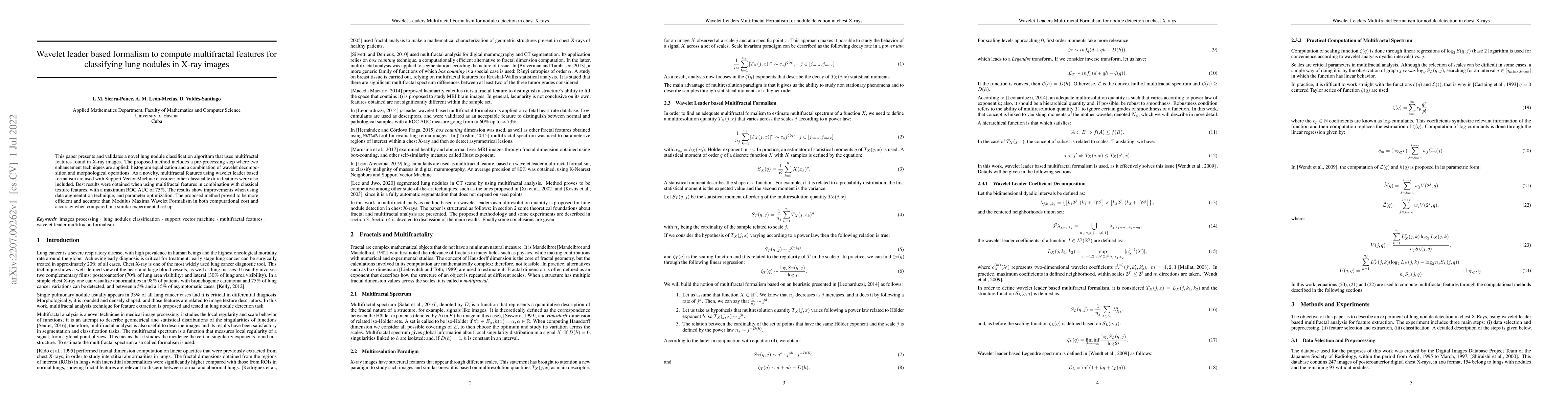
This paper presents and validates a novel lung nodule classification algorithm that uses multifractal features found in X-ray images. The proposed method includes a pre-processing step where two enhancement techniques are applied: histogram equalization and a combination of wavelet decomposition and morphological operations. As a novelty, multifractal features using wavelet leader based formalism are used with Support Vector Machine classifier; other classical texture features were also included. Best results were obtained when using multifractal features in combination with classical texture features, with a maximum ROC AUC of 75\%. The results show improvements when using data augmentation technique, and parameter optimization. The proposed method proved to be more efficient and accurate than Modulus Maxima Wavelet Formalism in both computational cost and accuracy when compared in a similar experimental set up.
AI Key Findings
Get AI-generated insights about this paper's methodology, results, significance, and more — seven facets brought into focus.
Impact
Paper Details
Authors
PDF Preview
Key Terms
Citation Network
Current paper (gray), citations (green), references (blue)
Display is limited for performance on very large graphs.
Discussion 0