Publication
Metrics
AI Quick Summary
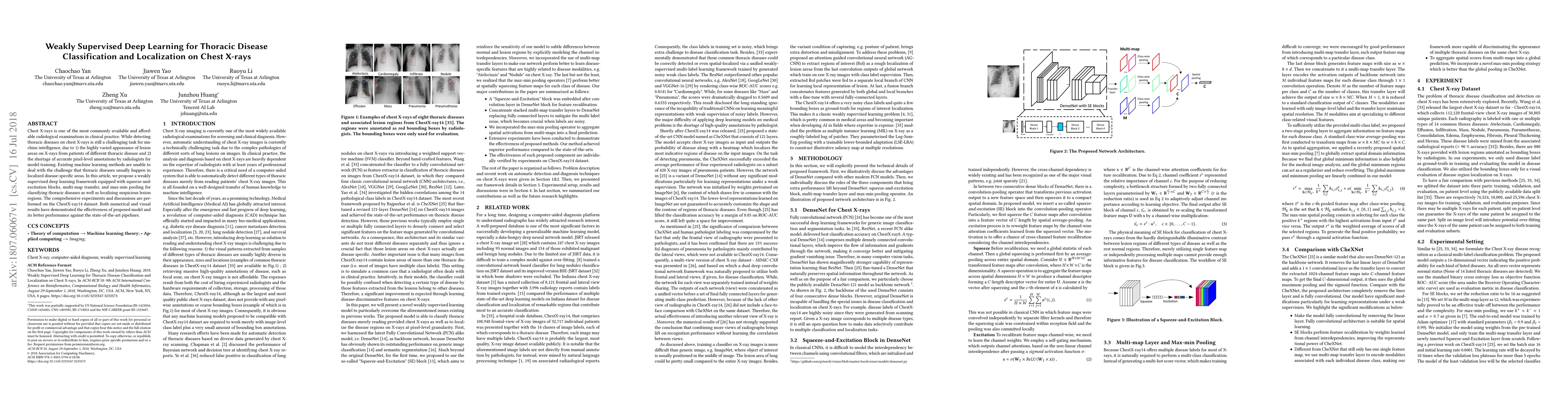
This paper proposes a weakly supervised deep learning framework for classifying thoracic diseases and localizing lesions on chest X-rays using squeeze-and-excitation blocks, multi-map transfer, and max-min pooling. Experiments on the ChestX-ray14 dataset show superior performance compared to state-of-the-art methods.
Paper Preview
Abstract
Chest X-rays is one of the most commonly available and affordable radiological examinations in clinical practice. While detecting thoracic diseases on chest X-rays is still a challenging task for machine intelligence, due to 1) the highly varied appearance of lesion areas on X-rays from patients of different thoracic disease and 2) the shortage of accurate pixel-level annotations by radiologists for model training. Existing machine learning methods are unable to deal with the challenge that thoracic diseases usually happen in localized disease-specific areas. In this article, we propose a weakly supervised deep learning framework equipped with squeeze-and-excitation blocks, multi-map transfer, and max-min pooling for classifying thoracic diseases as well as localizing suspicious lesion regions. The comprehensive experiments and discussions are performed on the ChestX-ray14 dataset. Both numerical and visual results have demonstrated the effectiveness of the proposed model and its better performance against the state-of-the-art pipelines.
AI Key Findings
Get AI-generated insights about this paper's methodology, results, significance, and more — seven facets brought into focus.
Impact
Paper Details
PDF Preview
Key Terms
Citation Network
Current paper (gray), citations (green), references (blue)
Display is limited for performance on very large graphs.
Discussion 0