Quick Summary:

A new weakly supervised object detection method using neural networks detects brain lesions in medical images without labeled examples, achieving results comparable to human agreement and outperforming other methods in some regions.

Summary

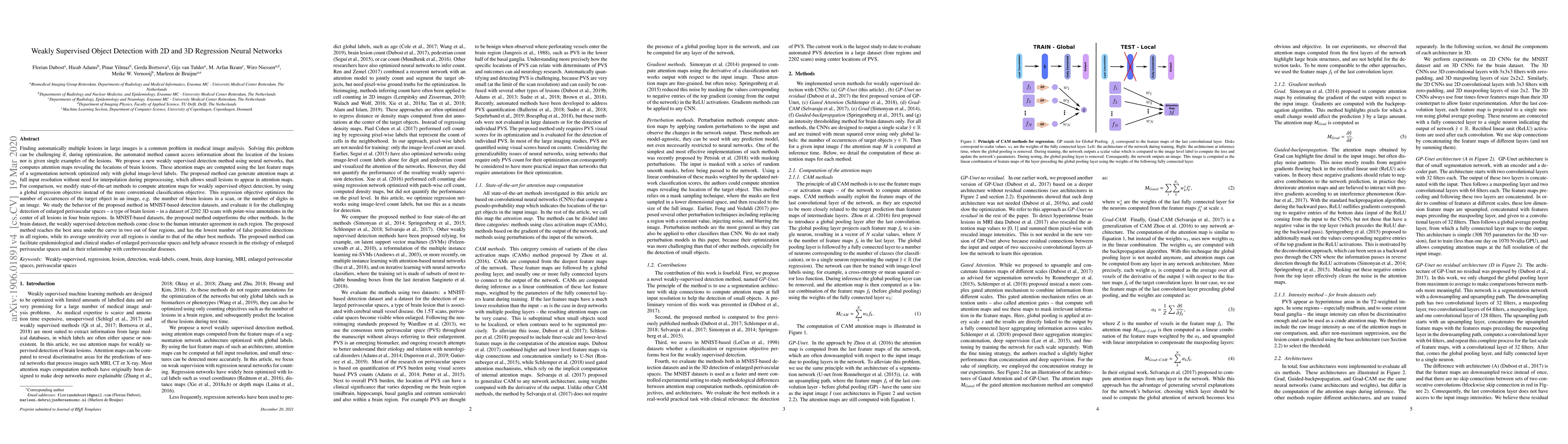
Finding automatically multiple lesions in large images is a common problem in medical image analysis. Solving this problem can be challenging if, during optimization, the automated method cannot access information about the location of the lesions nor is given single examples of the lesions. We propose a new weakly supervised detection method using neural networks, that computes attention maps revealing the locations of brain lesions. These attention maps are computed using the last feature maps of a segmentation network optimized only with global image-level labels. The proposed method can generate attention maps at full input resolution without need for interpolation during preprocessing, which allows small lesions to appear in attention maps. For comparison, we modify state-of-the-art methods to compute attention maps for weakly supervised object detection, by using a global regression objective instead of the more conventional classification objective. This regression objective optimizes the number of occurrences of the target object in an image, e.g. the number of brain lesions in a scan, or the number of digits in an image. We study the behavior of the proposed method in MNIST-based detection datasets, and evaluate it for the challenging detection of enlarged perivascular spaces - a type of brain lesion - in a dataset of 2202 3D scans with point-wise annotations in the center of all lesions in four brain regions. In the brain dataset, the weakly supervised detection methods come close to the human intrarater agreement in each region. The proposed method reaches the best area under the curve in two out of four regions, and has the lowest number of false positive detections in all regions, while its average sensitivity over all regions is similar to that of the other best methods. The proposed method can facilitate epidemiological and clinical studies of enlarged perivascular spaces.

AI Key Findings

Get AI-generated insights about this paper's methodology, results, and significance.

Paper Details

PDF Preview

Download PDF

Key Terms

brain (0.260)
regression (0.144)
proposed (0.115)
clinical (0.096)
feature (0.068)
studies (0.062)
propose (0.048)
study (0.041)

Citation Network

Current paper (gray), citations (green), references (blue)

Current Paper
Citations
References
Click to view
Title Authors Year Actions
Deep Learning Approaches for Medical Imaging Under Varying Degrees of Label Availability: A Comprehensive Survey
Siteng Ma, Honghui Du, Yu An, Jing Wang, Qinqin Wang, Haochang Wu, Aonghus Lawlor, Ruihai Dong
2025
View Paper
A Survey on Explainable Artificial Intelligence (XAI) Techniques for Visualizing Deep Learning Models in Medical Imaging
Deepshikha Bhati, Fnu Neha, Md Amiruzzaman
2024
View Paper
Deep Learning-Based Automated Measurement of Murine Bone Length in Radiographs
Ruichen Rong, Kristin Denton, Kevin W. Jin, Peiran Quan, Zhuoyu Wen, J. Kozlitina, Stephen Lyon, Aileen Wang, Carol A. Wise, Bruce Beutler, Donghan M. Yang, Qiwei Li, J. Rios, Guanghua Xiao
2024
View Paper
Systematic review and meta-analysis of automated methods for quantifying enlarged perivascular spaces in the brain
J. Waymont, M. C. V. Hernández, J. Bernal, R. D. Coello, Rosalind Brown, F. M. Chappell, L. Ballerini, Joanna M. Wardlaw
2024
View Paper
SE3D: A Framework For Saliency Method Evaluation In 3D Imaging
Mariusz Wi'sniewski, Loris Giulivi, Giacomo Boracchi
2024
View Paper
Perivascular space Identification Nnunet for Generalised Usage (PINGU)
Ben Sinclair, L. Vivash, Jasmine Moses, Miranda Lynch, William Pham, Karina Dorfman, Cassandra Marotta, Shaun Koh, Jacob Bunyamin, Ella Rowsthorn, Alex Jarema, Himashi Peiris, Zhaolin Chen, Sandy R. Shultz, David K Wright, Dexiao Kong, S. L. Naismith, Terence J. O'Brien, Meng Law
2024
View Paper
Evaluating the Potential of Metastatic Mediastinal Lymph Node Involvement in Non-Small Cell Lung Cancer Patients through Convolutional Neural Networks Analysis of Chest CT
A. P. Reprint, A. Shevtsov, Andrey O. Nefedov, Iaroslav D. Tominin, Piotr K. Yablonskii, Vladislav D. Tominin, P. Gavrilov, Vsevolod M. Malevanniy, Mariya E. Blokhina, Y. Esakov, Z. Tukvadze, V. Gombolevskiy, Yuriy Vasilev, Mariya Dugova, V. Kozlov, V. Chernina, E. Nalivkina, O. Omelyanskaya, M. Belyaev, Roman Reshetnikov, I. Blokhin
2024
View Paper
Beyond Pixel-Wise Supervision for Medical Image Segmentation: From Traditional Models to Foundation Models
Yuyan Shi, Jialu Ma, Jin Yang, Shasha Wang, Yichi Zhang
2024
View Paper
Score-Based Counterfactual Generation for Interpretable Medical Image Classification and Lesion Localization
Ke Wang, Zicong Chen, Mingjia Zhu, Zhetao Li, Jian Weng, Tianlong Gu
2024
View Paper
Weakly-supervised auto-encoder via energy regularization and soft multi-label learning on k labeled samples
Huiling Wang, Jun Sun, Xiaofeng Gu, Zunhao Hu, Chao Zhou
2024
View Paper
Attention-guided jaw bone lesion diagnosis in panoramic radiography using minimal labeling effort
Minseon Gwak, Jong Pil Yun, Ji Yun Lee, Sang-Sun Han, Poogyeon Park, Chena Lee
2024
View Paper
Overview of the Current Knowledge and Conventional MRI Characteristics of Peri- and Para-Vascular Spaces
M. Parillo, Federica Vaccarino, G. Di Gennaro, Sumeet Kumar, J. V. Van Goethem, B. Beomonte Zobel, C. C. Quattrocchi, Paul M Parizel, C. Mallio
2024
View Paper
Leveraging point annotations in segmentation learning with boundary loss
Eva Breznik, H. Kervadec, F. Malmberg, J. Kullberg, Haakan Ahlstrom, Marleen de Bruijne, R. Strand
2023
View Paper
Strengthening deep-learning models for intracranial hemorrhage detection: strongly annotated computed tomography images and model ensembles
Dong-Wan Kang, Gi-Hun Park, Wi-Sun Ryu, Dawid Schellingerhout, Museong Kim, Yong Soo Kim, Chan-Young Park, Keon-Joo Lee, Moon‐Ku Han, Han-Gil Jeong, Dong-Eog Kim
2023
View Paper
Atlas-Based Interpretable Age Prediction
Sophie Starck, Yadunandan Vivekanand Kini, J. Ritter, R. Braren, D. Rueckert, Tamara T. Mueller
2023
View Paper
Weakly Supervised Lesion Detection and Diagnosis for Breast Cancers With Partially Annotated Ultrasound Images
Jian Wang, Liang Qiao, Shichong Zhou, Jin Zhou, Jun Wang, Juncheng Li, Shihui Ying, Cai Chang, Jun Shi
2023
View Paper
Recent Advances in Explainable Artificial Intelligence for Magnetic Resonance Imaging
Jinzhao Qian, Hailong Li, Junqi Wang, Lili He
2023
View Paper
Fine-graded image classification network based on Faster MMAL-Net
Ke Han, Xiu Ji, Beimin Xie, Yong Wang, Shuai Gao
2023
View Paper
Label-Efficient Deep Learning in Medical Image Analysis: Challenges and Future Directions
Cheng Jin, Zhengrui Guo, Yi-Mou Lin, Luyang Luo, Hao Chen
2023
View Paper
Deep learning based identification of bone scintigraphies containing metastatic bone disease foci
A. Ibrahim, A. Vaidyanathan, S. Primakov, Flore Belmans, F. Bottari, T. Refaee, P. Lovinfosse, A. Jadoul, C. Derwael, F. Hertel, H. Woodruff, H. Zacho, Sean Walsh, W. Vos, M. Occhipinti, F. Hanin, P. Lambin, F. Mottaghy, R. Hustinx
2023
View Paper
ATCON: Attention Consistency for Vision Models
Ali Mirzazadeh, Florian Dubost, M. Pike, Krish Maniar, Max Zuo, Christopher Lee-Messer, D. Rubin
2022
View Paper
Determinants of Perivascular Spaces in the General Population
T. Evans, M. Knol, P. Schwingenschuh, K. Wittfeld, S. Hilal, M. A. Ikram, Florian Dubost, Kimberlin M H van Wijnen, P. Katschnig, Pinar Yilmaz, Marleen de Bruijne, M. Habes, Christopher Chen, Sönke Langer, H. Völzke, M. K. Ikram, H. Grabe, Reinhold Schmidt, H. Adams, Meike W. Vernooij
2022
View Paper
Assessment of perivascular space filtering methods using a three-dimensional computational model.
J. Bernal, M. Valdés-Hernández, J. Escudero, Roberto Duarte, L. Ballerini, M. Bastin, I. Deary, M. Thrippleton, R. Touyz, J. Wardlaw
2022
View Paper
Attention-Based Dynamic Subspace Learners for Medical Image Analysis
V. SukeshAdiga, J. Dolz, H. Lombaert
2022
View Paper
Microscopy analysis neural network to solve detection, enumeration and segmentation from image-level annotations
Anthony Bilodeau, Constantin V. L. Delmas, M. Parent, P. De Koninck, A. Durand, Flavie Lavoie-Cardinal
2022
View Paper
Segmentation-Consistent Probabilistic Lesion Counting
J. Schroeter, Chelsea Myers-Colet, D. Arnold, T. Arbel
2022
View Paper
Computer-aided extraction of select MRI markers of cerebral small vessel disease: A systematic review
Jiyang Jiang, Dadong Wang, Yang Song, P. Sachdev, W. Wen
2022
View Paper
Breast Tumor Classification Based on MRI-US Images by Disentangling Modality Features
Mengyun Qiao, Chengcheng Liu, Zeju Li, Jin Zhou, Qin Xiao, Shichong Zhou, Cai Chang, G. Yajia, Yi Guo, Yuanyuan Wang
2022
View Paper
Arthroscope Localization in 3D Ultrasound Volumes Using Weakly Supervised Deep Learning
Jeroen M. A. van der Burgt, S. Camps, M. Antico, G. Carneiro, D. Fontanarosa
2021
View Paper
Explainable artificial intelligence (XAI) in deep learning-based medical image analysis
Bas H. M. van der Velden, Hugo J. Kuijf, K. Gilhuijs, M. Viergever
2021
View Paper
DLSE-Net: A robust weakly supervised network for fabric defect detection
Zhoufeng Liu, Zhaochen Huo, Chunlei Li, Yan Dong, Bicao Li
2021
View Paper
Weakly Supervised Object Localization and Detection: A Survey
Dingwen Zhang, Junwei Han, Gong Cheng, Ming-Hsuan Yang
2021
View Paper
Information Bottleneck Attribution for Visual Explanations of Diagnosis and Prognosis
Ugur Demir, I. Irmakci, E. Keles, A. Topçu, Ziyue Xu, C. Spampinato, S. Jambawalikar, E. Turkbey, B. Turkbey, Ulas Bagci
2021
View Paper
3D Segmentation of Perivascular Spaces on T1-Weighted 3 Tesla MR Images With a Convolutional Autoencoder and a U-Shaped Neural Network
P. Boutinaud, A. Tsuchida, Alexandre Laurent, Filipa Adonias, Zahra Hanifehlou, Victor Nozais, Violaine Verrecchia, L. Lampe, Junyi Zhang, Yicheng Zhu, C. Tzourio, B. Mazoyer, M. Joliot
2020
View Paper
Hierarchical class grouping with orthogonal constraint for class activation map generation
Fanman Meng, Huang Kaixu, Hongliang Li, Shuaipu Chen, Q. Wu, K. Ngan
2020
View Paper
MICRA-Net: MICRoscopy Analysis Neural Network to solve detection, classification, and segmentation from a single simple auxiliary task
Anthony Bilodeau, Constantin V. L. Delmas, M. Parent, P. De Koninck, A. Durand, Flavie Lavoie-Cardinal
2020
View Paper
Bathymetric Inversion and Uncertainty Estimation from Synthetic Surf-Zone Imagery with Machine Learning
Adam Collins, K. Brodie, A. Bak, T. Hesser, M. Farthing, Jonghyun Lee, J. W. Long
2020
View Paper
ExplAIn: Explanatory Artificial Intelligence for Diabetic Retinopathy Diagnosis
G. Quellec, Hassan Al Hajj, M. Lamard, Pierre-Henri Conze, P. Massin, B. Cochener
2020
View Paper
Automated Quantification of Enlarged Perivascular Spaces in Clinical Brain MRI across Sites
Florian Dubost, M. Dünnwald, Denver Huff, V. Scheumann, F. Schreiber, M. Vernooij, W. Niessen, M. Skalej, S. Schreiber, S. Oeltze-Jafra, Marleen de Bruijne
2019
View Paper
Computer Methods for Automatic Locomotion and Gesture Tracking in Mice and Small Animals for Neuroscience Applications: A Survey
W. Abbas, D. Masip
2019
View Paper
Weakly Supervised Virus Capsid Detection with Image-Level Annotations in Electron Microscopy Images
H. Kniesel, Leon Sick, Tristan Payer, T. Bergner, K. S. Devan, C. Read, Paul Walther, Timo Ropinski, P. Hermosilla
2024
View Paper
Determinants of Perivascular Spaces in the General Population A Pooled Cohort Analysis of Individual Participant Data
T. Evans, M. Knol, P. Schwingenschuh, K. Wittfeld, S. Hilal, M. Ikram, Florian Dubost, Kimberlin M H van Wijnen, P. Katschnig, P. Yilmaz, Marleen de Bruijne, M. Habes, Christopher P L H Chen, Sönke Langer, H. Völzke, M. Ikram, H. Grabe, R. Schmidt, H. Adams, M. Vernooij
View Paper
Medical Image Analysis
Wenhui Lei, Wei Xu, Kang Li, Xiaofan Zhang, Shaoting Zhang
View Paper

Comments (0)

This website uses cookies to ensure you get the best experience. Learn more