What Can Machine Vision Do for Lymphatic Histopathology Image Analysis: A Comprehensive Review
Publication
Metrics
AI Quick Summary
This paper reviews advancements in machine vision for analyzing lymphatic histopathology images, focusing on segmentation, classification, and detection using deep learning. It highlights current methods and proposes future directions for improved diagnostic accuracy.
Paper Preview
Abstract
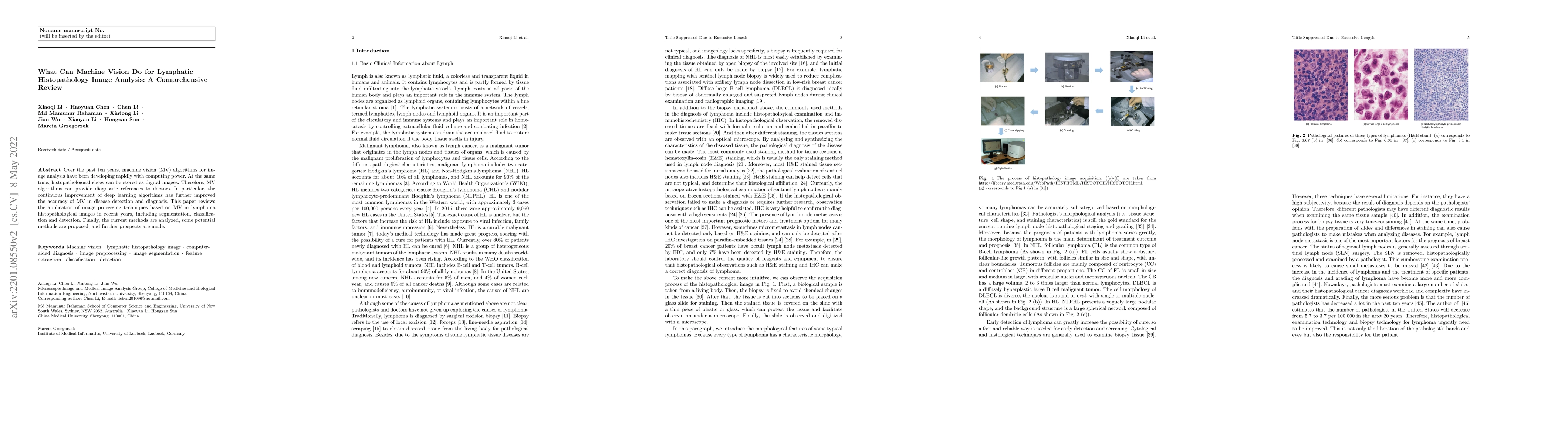
In the past ten years, the computing power of machine vision (MV) has been continuously improved, and image analysis algorithms have developed rapidly. At the same time, histopathological slices can be stored as digital images. Therefore, MV algorithms can provide doctors with diagnostic references. In particular, the continuous improvement of deep learning algorithms has further improved the accuracy of MV in disease detection and diagnosis. This paper reviews the applications of image processing technology based on MV in lymphoma histopathological images in recent years, including segmentation, classification and detection. Finally, the current methods are analyzed, some more potential methods are proposed, and further prospects are made.
AI Key Findings
Get AI-generated insights about this paper's methodology, results, significance, and more — seven facets brought into focus.
Impact
Paper Details
Authors
PDF Preview
Key Terms
Citation Network
Current paper (gray), citations (green), references (blue)
Display is limited for performance on very large graphs.
Discussion 0