When are Diffusion Priors Helpful in Sparse Reconstruction? A Study with Sparse-view CT

Source: ArXiv

AI Quick Summary

This study examines the effectiveness of diffusion priors in sparse-view CT reconstruction, finding that while they can capture significant detail with few observations, they perform worse than classical priors when sufficient data is available. The research emphasizes the need for further investigation into the use of diffusion models in clinical settings.

Share: Share on X LinkedIn

Summary

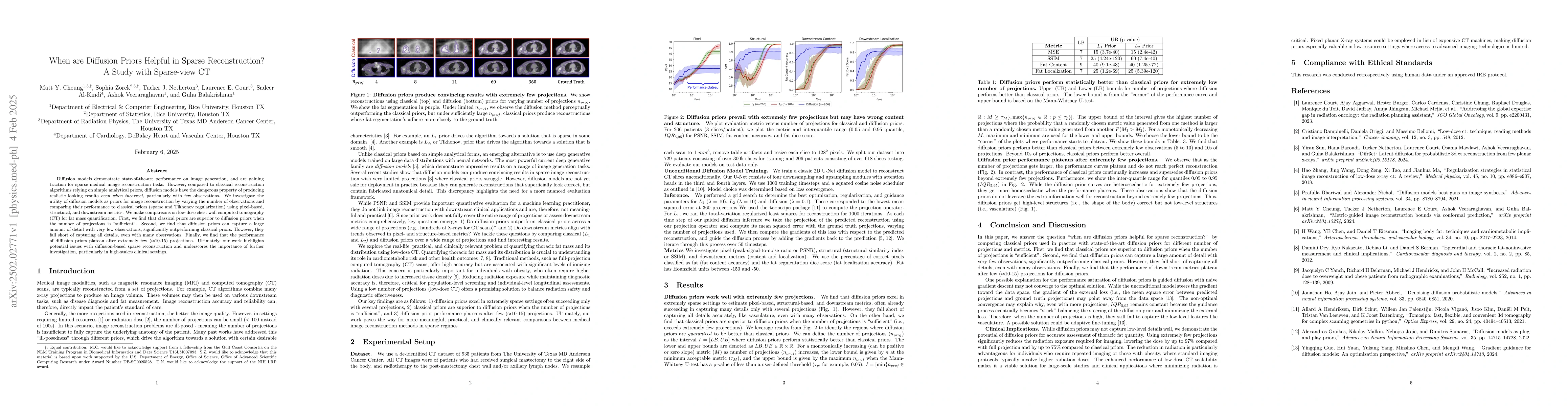
Diffusion models demonstrate state-of-the-art performance on image generation, and are gaining traction for sparse medical image reconstruction tasks. However, compared to classical reconstruction algorithms relying on simple analytical priors, diffusion models have the dangerous property of producing realistic looking results \emph{even when incorrect}, particularly with few observations. We investigate the utility of diffusion models as priors for image reconstruction by varying the number of observations and comparing their performance to classical priors (sparse and Tikhonov regularization) using pixel-based, structural, and downstream metrics. We make comparisons on low-dose chest wall computed tomography (CT) for fat mass quantification. First, we find that classical priors are superior to diffusion priors when the number of projections is ``sufficient''. Second, we find that diffusion priors can capture a large amount of detail with very few observations, significantly outperforming classical priors. However, they fall short of capturing all details, even with many observations. Finally, we find that the performance of diffusion priors plateau after extremely few ($\approx$10-15) projections. Ultimately, our work highlights potential issues with diffusion-based sparse reconstruction and underscores the importance of further investigation, particularly in high-stakes clinical settings.

AI Key Findings

Generated Jun 12, 2025

Methodology

The study compares classical priors (L1 and L2) with diffusion priors for sparse image reconstruction in low-dose CT scans using pixel-based, structural, and downstream metrics. A dataset of 935 CT scans from The University of Texas MD Anderson Cancer Center was used, with 729 patients for training and 206 for testing. An unconditional diffusion model was trained using a 2D U-Net architecture, and grid search was performed to determine the best optimization, regularization, and guidance parameters.

Key Results

  • Classical priors outperform diffusion priors when the number of projections is sufficient.
  • Diffusion priors capture a large amount of detail with very few observations, significantly outperforming classical priors in extremely sparse settings.
  • Diffusion prior performance plateaus after approximately 10-15 projections.
  • Diffusion priors perform better than classical priors between extremely few observations (5 to 10) and around 100s of projections.
  • Diffusion priors do not leverage extra information well for reconstruction beyond extremely few projections, getting high-level structures correct but not low-level structures like vasculature.

Significance

This research highlights potential issues with diffusion-based sparse reconstruction and underscores the importance of further investigation, particularly in high-stakes clinical settings. It demonstrates the potential of diffusion priors for accurate assessment of thoracic fat quantity while significantly reducing radiation exposure required for imaging, making low-dose CT a viable solution for large-scale studies and clinical applications where minimizing radiation is critical.

Technical Contribution

The study presents a comprehensive comparison of classical and diffusion priors for sparse image reconstruction in low-dose CT scans, evaluating their performance using various metrics and demonstrating the potential benefits and limitations of diffusion priors in this context.

Novelty

This work is novel in its systematic investigation of diffusion priors for sparse reconstruction tasks in medical imaging, comparing them against classical priors and evaluating their performance using pixel-based, structural, and downstream metrics. The research also emphasizes the importance of downstream clinical metrics and highlights the need for more nuanced evaluation frameworks in this domain.

Limitations

  • Diffusion priors may not capture low-level details well, such as vasculature, even with many observations.
  • The performance of diffusion priors plateaus after a relatively small number of projections (around 10-15).

Future Work

  • Explore adaptive fine-tuning to improve convergence and capture low-level features better.
  • Investigate the use of diffusion priors in other medical imaging modalities and applications.

Paper Details

PDF Preview

Download PDF

Citation Network

Current paper (gray), citations (green), references (blue)

Display is limited for performance on very large graphs.

Current Paper
Citations
References
Click to view

No citations found for this paper.

Comments (0)

This website uses cookies to ensure you get the best experience. Learn more