Authors
Summary
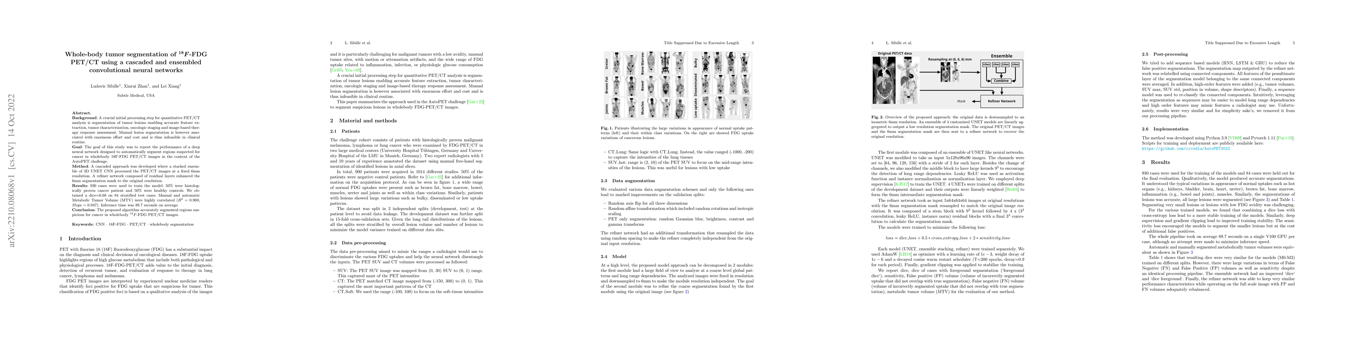
Background: A crucial initial processing step for quantitative PET/CT analysis is the segmentation of tumor lesions enabling accurate feature ex-traction, tumor characterization, oncologic staging, and image-based therapy response assessment. Manual lesion segmentation is however associated with enormous effort and cost and is thus infeasible in clinical routine. Goal: The goal of this study was to report the performance of a deep neural network designed to automatically segment regions suspected of cancer in whole-body 18F-FDG PET/CT images in the context of the AutoPET challenge. Method: A cascaded approach was developed where a stacked ensemble of 3D UNET CNN processed the PET/CT images at a fixed 6mm resolution. A refiner network composed of residual layers enhanced the 6mm segmentation mask to the original resolution. Results: 930 cases were used to train the model. 50% were histologically proven cancer patients and 50% were healthy controls. We obtained a dice=0.68 on 84 stratified test cases. Manual and automatic Metabolic Tumor Volume (MTV) were highly correlated (R2 = 0.969,Slope = 0.947). Inference time was 89.7 seconds on average. Conclusion: The proposed algorithm accurately segmented regions suspicious for cancer in whole-body 18F -FDG PET/CT images.
AI Key Findings
Get AI-generated insights about this paper's methodology, results, and significance.
Paper Details
PDF Preview
Key Terms
Citation Network
Current paper (gray), citations (green), references (blue)
Display is limited for performance on very large graphs.
Similar Papers
Found 4 papersAutomated Lesion Segmentation in Whole-Body FDG-PET/CT with Multi-modality Deep Neural Networks
Satoshi Kondo, Satoshi Kasai
Whole-Body Lesion Segmentation in 18F-FDG PET/CT
Zheng Zhang, Yuhang Shi, Jia Zhang et al.
Tumor Thrombus Associated With Locally Recurrent Osteosarcoma on [18F] FDG PET/CT.
Belge Bilgin, Gokce, Bilgin, Cem, Powell, Garret M et al.
| Title | Authors | Year | Actions |
|---|
Comments (0)