Summary

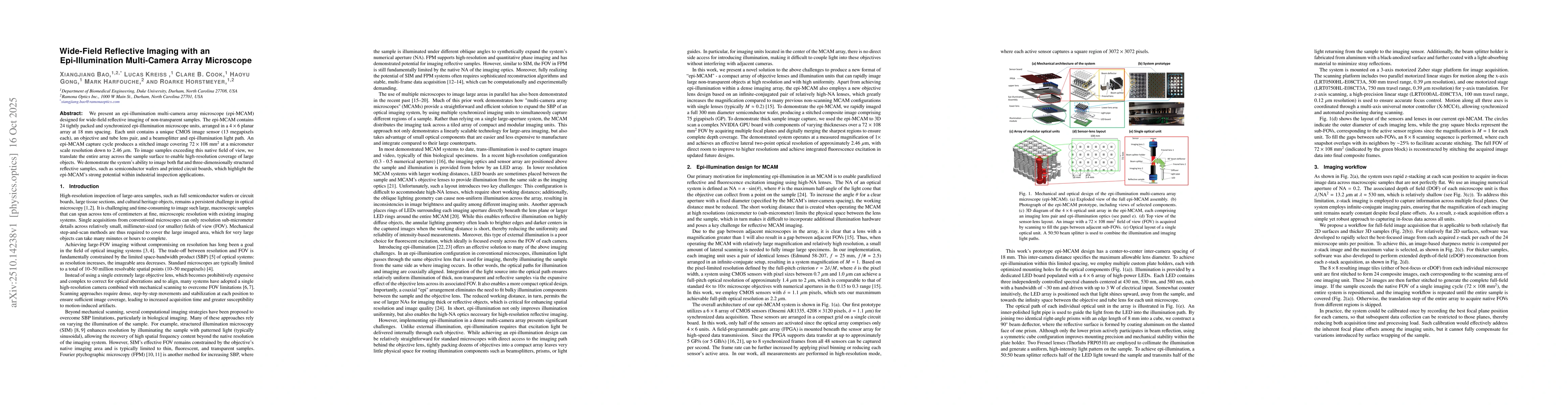
We present an epi-illumination multi-camera array microscope (epi-MCAM) designed for wide-field reflective imaging of non-transparent samples. The epi-MCAM contains 24 tightly packed and synchronized epi-illumination microscope units, arranged in a $4 \times 6$ planar array at 18 mm spacing. Each unit contains a unique CMOS image sensor (13 megapixels each), an objective and tube lens pair, and a beamsplitter and epi-illumination light path. An epi-MCAM capture cycle produces a stitched image covering $72 \times 108~\mathrm{mm}^2$ at a micrometer scale resolution down to 2.46 $\mu$m. To image samples exceeding this native field of view, we translate the entire array across the sample surface to enable high-resolution coverage of large objects. We demonstrate the system's ability to image both flat and three-dimensionally structured reflective samples, such as semiconductor wafers and printed circuit boards, which highlight the epi-MCAM's strong potential within industrial inspection applications.

AI Key Findings

Generated Oct 20, 2025

Methodology

The study presents an epi-illumination multi-camera array microscope system that combines structured illumination and multi-camera imaging techniques to achieve high-resolution, large-field-of-view imaging. It employs a flatbed scanner for capturing gigapixel images and utilizes Fourier ptychographic microscopy principles for phase retrieval and resolution enhancement.

Key Results

  • Achieved 0.5 gigapixel resolution with submicron spatial accuracy across centimeter-scale fields of view
  • Demonstrated multi-scale imaging capabilities by integrating structured illumination with multi-camera array technology
  • Enabled non-contact, high-resolution imaging of biological specimens with improved phase contrast

Significance

This research advances optical microscopy by providing a cost-effective solution for high-resolution, large-field-of-view imaging. The technology has potential applications in biomedical imaging, digital pathology, and materials science for non-invasive, high-throughput analysis.

Technical Contribution

The technical contribution lies in the integration of structured illumination with multi-camera array systems, combined with Fourier ptychographic microscopy principles, to achieve unprecedented resolution and field of view in a single imaging platform.

Novelty

This work introduces a novel hybrid imaging approach that combines epi-illumination techniques with multi-camera array systems, offering significant improvements in resolution and field of view compared to traditional microscopy methods.

Limitations

  • Limited to flat sample geometries due to the flatbed scanner configuration
  • Requires complex computational processing for image reconstruction
  • May have reduced depth of field compared to traditional microscopes

Future Work

  • Development of 3D imaging capabilities with extended depth of field
  • Integration with machine learning algorithms for automated defect detection
  • Adaptation for in vivo biological imaging applications
  • Optimization for real-time imaging with reduced computational load

Paper Details

PDF Preview

Download PDF

Comments (0)

This website uses cookies to ensure you get the best experience. Learn more