Publication
Metrics
AI Quick Summary
This paper presents a wide-field strain imaging technique using optically detected magnetic resonance of NV centers in polycrystalline diamond, revealing a heterogeneous crystalline environment with long spin coherence times and high sensitivity for 3D strain imaging. The method leverages the preferential alignment of NV centers for diffraction-limited resolution across a broad field of view.
Paper Preview
Abstract
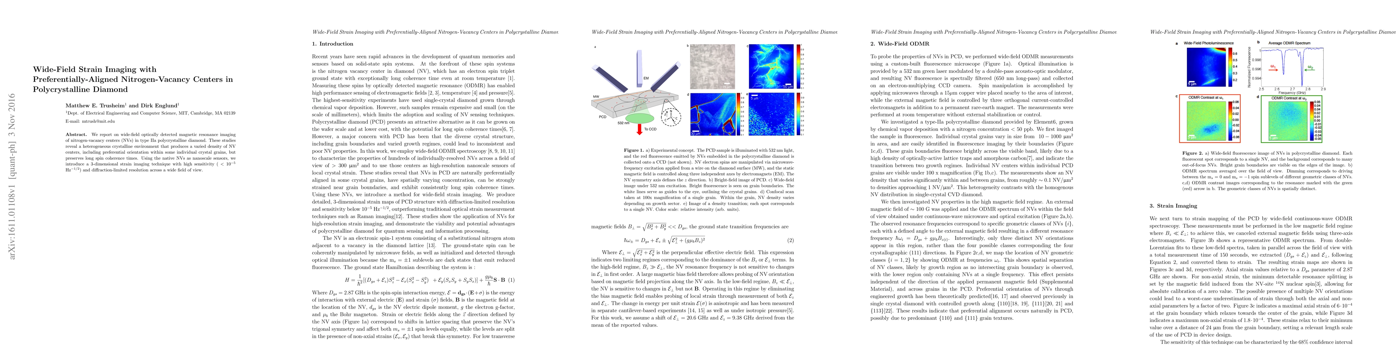
We report on wide-field optically detected magnetic resonance imaging of nitrogen-vacancy centers (NVs) in type IIa polycrystalline diamond. These studies reveal a heterogeneous crystalline environment that produces a varied density of NV centers, including preferential orientation within some individual crystal grains, but preserves long spin coherence times. Using the native NVs as nanoscale sensors, we introduce a 3-dimensional strain imaging technique with high sensitivity ( $< 10^{-5}$ Hz$^{-1/2}$) and diffraction-limited resolution across a wide field of view.
AI Key Findings
Get AI-generated insights about this paper's methodology, results, significance, and more — seven facets brought into focus.
Impact
Paper Details
PDF Preview
Key Terms
Citation Network
Current paper (gray), citations (green), references (blue)
Display is limited for performance on very large graphs.
Discussion 0