Wound Tissue Segmentation in Diabetic Foot Ulcer Images Using Deep Learning: A Pilot Study
Publication
Metrics
AI Quick Summary
This paper presents a pilot study on segmenting diabetic foot ulcer tissues using a hybrid deep learning model combining Mix Transformer and CNN with a P-scSE module. The study introduces a new DFUTissue dataset to evaluate segmentation algorithms, employing both supervised and semi-supervised learning to improve performance. The hybrid model achieves a Dice Similarity Coefficient of 87.64% in the semi-supervised phase, outperforming state-of-the-art methods.
Paper Preview
Abstract
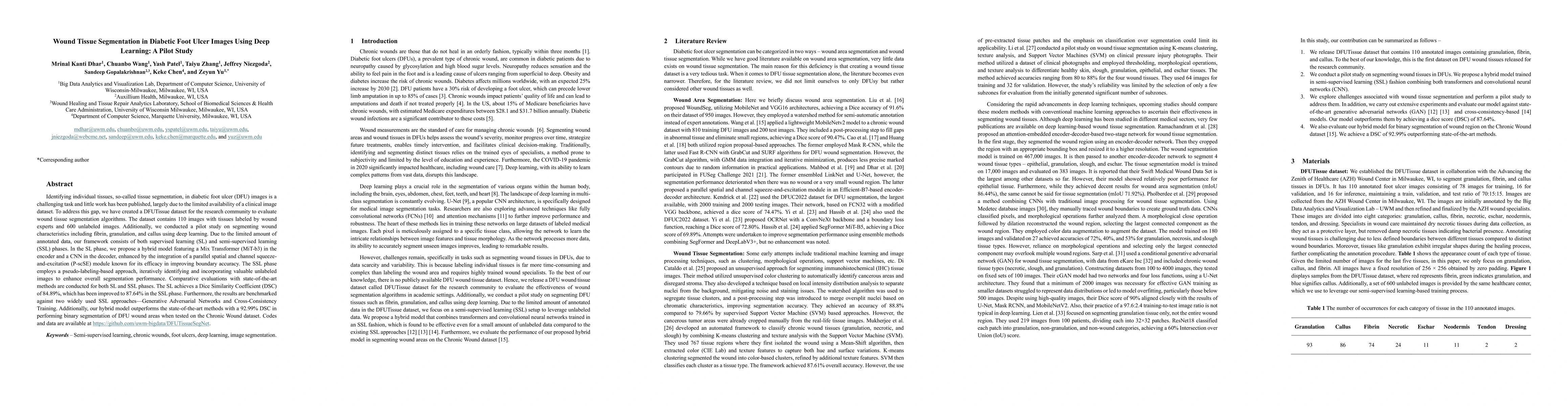
Identifying individual tissues, so-called tissue segmentation, in diabetic foot ulcer (DFU) images is a challenging task and little work has been published, largely due to the limited availability of a clinical image dataset. To address this gap, we have created a DFUTissue dataset for the research community to evaluate wound tissue segmentation algorithms. The dataset contains 110 images with tissues labeled by wound experts and 600 unlabeled images. Additionally, we conducted a pilot study on segmenting wound characteristics including fibrin, granulation, and callus using deep learning. Due to the limited amount of annotated data, our framework consists of both supervised learning (SL) and semi-supervised learning (SSL) phases. In the SL phase, we propose a hybrid model featuring a Mix Transformer (MiT-b3) in the encoder and a CNN in the decoder, enhanced by the integration of a parallel spatial and channel squeeze-and-excitation (P-scSE) module known for its efficacy in improving boundary accuracy. The SSL phase employs a pseudo-labeling-based approach, iteratively identifying and incorporating valuable unlabeled images to enhance overall segmentation performance. Comparative evaluations with state-of-the-art methods are conducted for both SL and SSL phases. The SL achieves a Dice Similarity Coefficient (DSC) of 84.89%, which has been improved to 87.64% in the SSL phase. Furthermore, the results are benchmarked against two widely used SSL approaches: Generative Adversarial Networks and Cross-Consistency Training. Additionally, our hybrid model outperforms the state-of-the-art methods with a 92.99% DSC in performing binary segmentation of DFU wound areas when tested on the Chronic Wound dataset. Codes and data are available at https://github.com/uwm-bigdata/DFUTissueSegNet.
AI Key Findings
Get AI-generated insights about this paper's methodology, results, significance, and more — seven facets brought into focus.
Impact
Authors
PDF Preview
Key Terms
Citation Network
Current paper (gray), citations (green), references (blue)
Display is limited for performance on very large graphs.
Discussion 0