Publication
Metrics
AI Quick Summary
This study synthesizes Cu2+ doped Zn1-xCuxFe2O4 spinel nanoparticles using a sol-gel method and analyzes their crystal structure via X-ray diffraction. The Williamson-Hall plot method reveals the effects of crystallite size and lattice strain on peak broadening, showing a reduction in lattice constant and unit cell volume with increased Cu2+ doping.
Paper Preview
Abstract
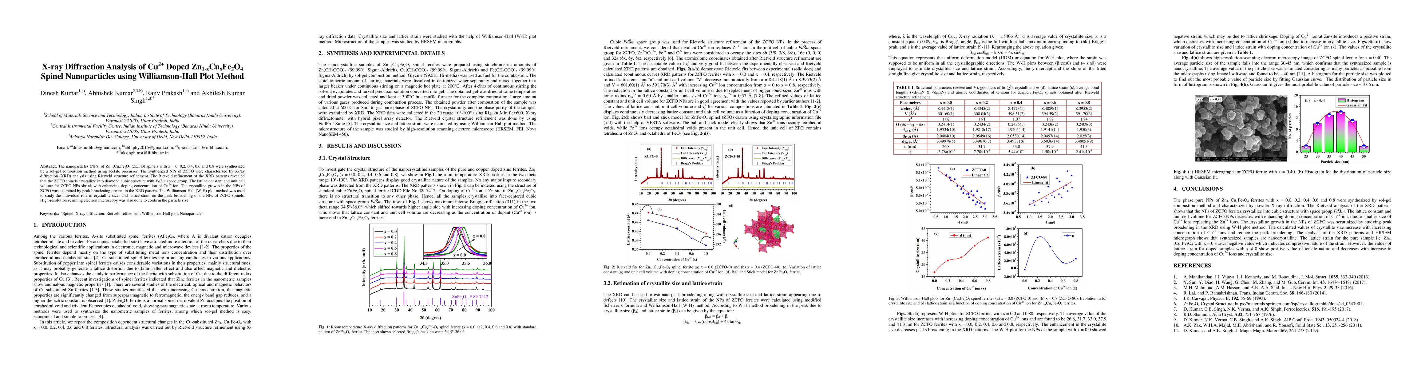
The nanoparticles (NPs) of Zn1-xCuxFe2O4 (ZCFO) spinels with x = 0, 0.2, 0.4, 0.6 and 0.8 were synthesized by a sol-gel combustion method using acetate precursor. The NPs of ZCFO were prepared by following calcination process at 600C for 8hrs. The synthesized NPs of ZCFO were characterized by X-ray diffraction (XRD) analysis using Rietveld refinement. The Rietveld refinement of the XRD patterns revealed that the ZCFO spinels crystallize into single diamond cubic structure with Fd-3m space group. The lattice constant and unit cell volume for ZCFO NPs shrink with enhancing doping concentration of Cu2+ ion. The crystalline growth in the NPs of ZCFO was examined by peak broadening present in the XRD pattern. The Williamson-Hall (W-H) plot method were used to study the individual involvements of crystallite sizes and lattice strain on the peak broadening of the NPs of ZCFO spinels. Whereas, particle size of the ZCFO sample with x = 0.40 was estimated by high-resolution scanning electron microscopy micrographs
AI Key Findings
Get AI-generated insights about this paper's methodology, results, significance, and more — seven facets brought into focus.
Impact
Paper Details
PDF Preview
Key Terms
Citation Network
Current paper (gray), citations (green), references (blue)
Display is limited for performance on very large graphs.
Discussion 0