01
MethodologyHow they did it
Micro scanning X-ray diffraction is used to characterize stress in suspended structures at sub micron scales
This research utilizes micro scanning X-ray diffraction to characterize stress in MEMS structures at sub-micron scales, specifically examining a bilayer cantilever and a boron-doped silicon bridge, with results validated by numerical simulations.
This research utilizes micro scanning X-ray diffraction to characterize stress in MEMS structures at sub-micron scales, specifically examining a bilayer cantilever and a boron-doped silicon bridge, with results validated by numerical simulations.
Micro scanning X-ray diffraction is used to characterize stress in suspended structures at sub micron scales More in Methodology →
The gold film gives a diffraction signal for both the white and monochromatic x-ray beam. — The array has been then imaged using gold fluorescence on fig. 1 (b). This allows for precisely calibrating the x-y sample stage. More in Key Results →
This research is important because it uses a promising technique that allows stress characterization in complex structures at sub micron scales More in Significance →
The polysilicon layer is almost stress free. — Grain boundary conditions used in modeling need to be improved. More in Limitations →
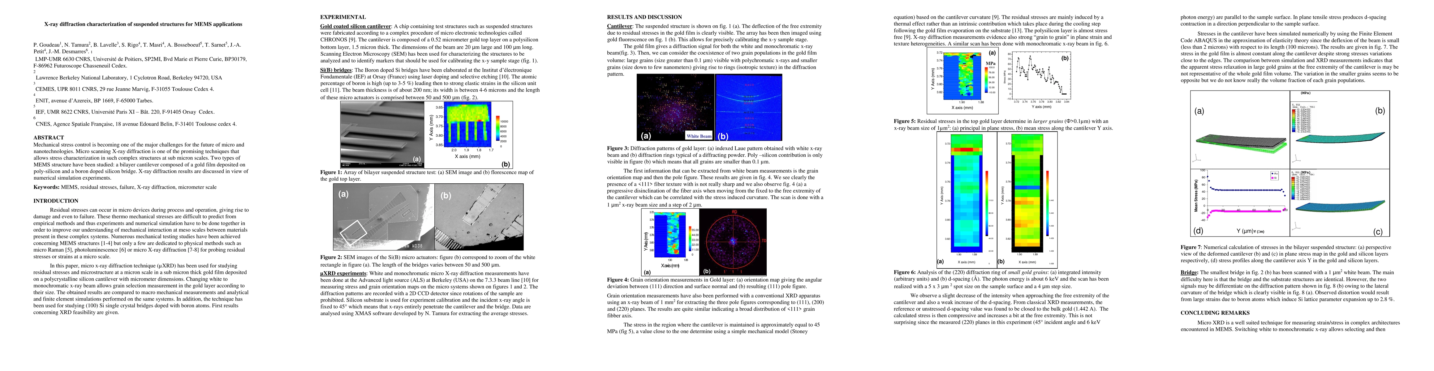
Mechanical stress control is becoming one of the major challenges for the future of micro and nanotechnologies. Micro scanning X-ray diffraction is one of the promising techniques that allows stress characterization in such complex structures at sub micron scales. Two types of MEMS structure have been studied: a bilayer cantilever composed of a gold film deposited on poly-silicon and a boron doped silicon bridge. X-ray diffraction results are discussed in view of numerical simulation experiments.
Seven facets of this paper, analysed and brought into focus by AI.
This research is important because it uses a promising technique that allows stress characterization in complex structures at sub micron scales
Micro scanning X-ray diffraction is used to characterize stress in suspended structures at sub micron scales
This research is important because it uses a promising technique that allows stress characterization in complex structures at sub micron scales
Numerical simulation experiments were performed using the Finite Element Code ABAQUS in the approximation of elasticity theory.
Micro XRD is a well suited technique for measuring strain/stress in complex architectures encountered in MEMS.
Current paper (gray), citations (green), references (blue)
Display is limited for performance on very large graphs.
Discussion 0