Publication
Metrics
AI Quick Summary
This paper presents a diffraction microscopy technique using refractive optics to study crystal structural variations, achieving micrometer resolution with a setup integrated into a 6-circle diffractometer at the ESRF beamline, adaptable for sub-micrometer resolution.
Paper Preview
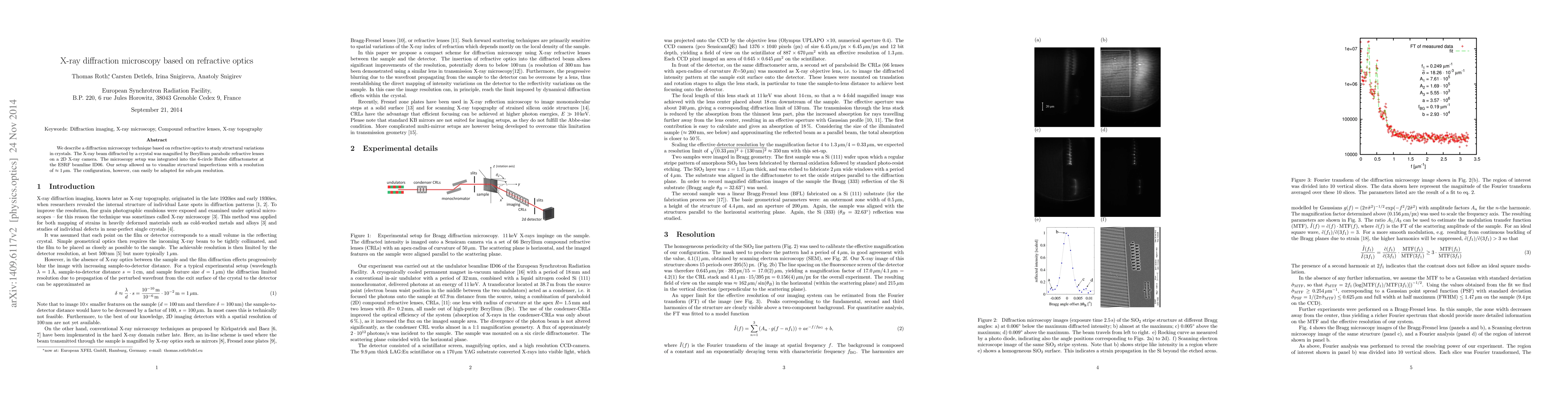
Abstract
We describe a diffraction microscopy technique based on refractive optics to study structural variations in crystals. The X-ray beam diffracted by a crystal was magnified by beryllium parabolic refractive lenses on a 2D X-ray camera. The microscopy setup was integrated into the 6-circle Huber diffractometer at the ESRF beamline ID06. Our setup allowed us to visualize structural imperfections with a resolution of approximately 1 micrometer. The configuration, however, can easily be adapted for sub-micrometer resolution.
AI Key Findings
Get AI-generated insights about this paper's methodology, results, significance, and more — seven facets brought into focus.
Impact
Paper Details
PDF Preview
Key Terms
Citation Network
Current paper (gray), citations (green), references (blue)
Display is limited for performance on very large graphs.
Discussion 0