Publication
Metrics
AI Quick Summary
This study introduces "X-ray dissectography" to enhance lung nodule detection by digitally separating lung structures from radiographic projections, thereby improving precision by over 20% compared to traditional methods. The proposed collaborative detection network localizes nodules in both 2D and 3D spaces, potentially revolutionizing chest X-ray imaging protocols.
Paper Preview
Abstract
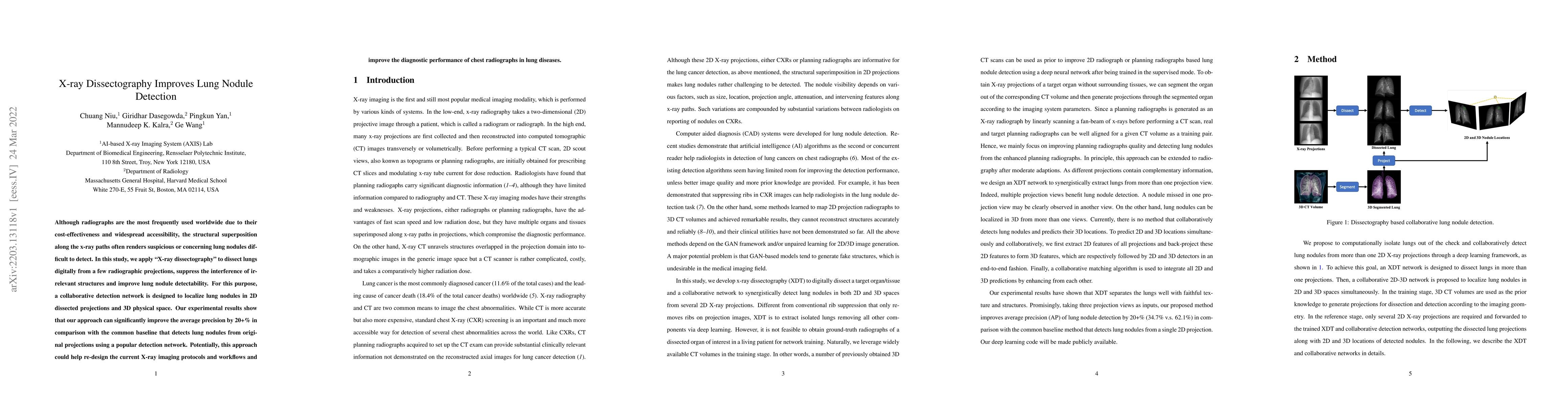
Although radiographs are the most frequently used worldwide due to their cost-effectiveness and widespread accessibility, the structural superposition along the x-ray paths often renders suspicious or concerning lung nodules difficult to detect. In this study, we apply "X-ray dissectography" to dissect lungs digitally from a few radiographic projections, suppress the interference of irrelevant structures, and improve lung nodule detectability. For this purpose, a collaborative detection network is designed to localize lung nodules in 2D dissected projections and 3D physical space. Our experimental results show that our approach can significantly improve the average precision by 20+% in comparison with the common baseline that detects lung nodules from original projections using a popular detection network. Potentially, this approach could help re-design the current X-ray imaging protocols and workflows and improve the diagnostic performance of chest radiographs in lung diseases.
AI Key Findings
Get AI-generated insights about this paper's methodology, results, significance, and more — seven facets brought into focus.
Impact
Paper Details
Authors
PDF Preview
Key Terms
Citation Network
Current paper (gray), citations (green), references (blue)
Display is limited for performance on very large graphs.
Discussion 0