X-ray fluorescence computed tomography (XFCT) imaging with a superfine pencil beam x-ray source
Publication
Metrics
AI Quick Summary
This paper presents a benchtop X-ray fluorescence computed tomography (XFCT) imaging system utilizing a superfine pencil beam x-ray source and an L1 regularization algorithm for improved molecular sensitivity and spatial resolution. The system's performance was evaluated using GATE simulations and compared to other reconstruction algorithms, showing promising results for future experimental testing.
Paper Preview
Abstract
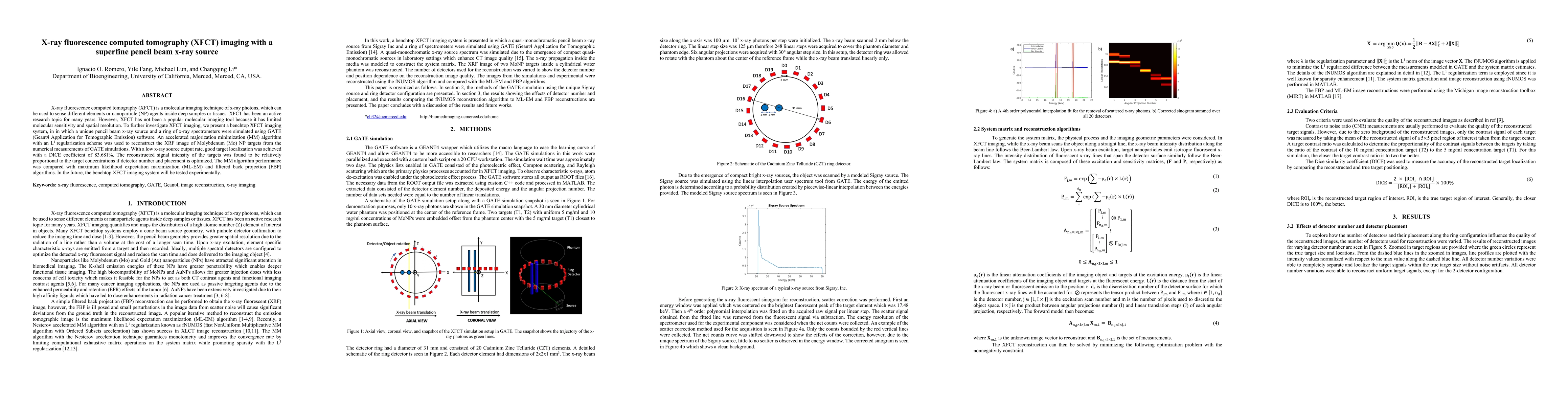
X-ray fluorescence computed tomography (XFCT) is a molecular imaging technique of x-ray photons, which can be used to sense different elements or nanoparticle (NP) agents inside deep samples or tissues. XFCT has been an active research topic for many years. However, XFCT has not been a popular molecular imaging tool because it has limited molecular sensitivity and spatial resolution. To further investigate XFCT imaging, we present a benchtop XFCT imaging system, in in which a unique pencil beam x-ray source and a ring of x-ray spectrometers were simulated using GATE (Geant4 Application for Tomographic Emission) software. An accelerated majorization minimization (MM) algorithm with an L1 regularization scheme was used to reconstruct the XRF image of Molybdenum (Mo) NP targets from the numerical measurements of GATE simulations. With a low x-ray source output rate, good target localization was achieved with a DICE coefficient of 83.681%. The reconstructed signal intensity of the targets was found to be relatively proportional to the target concentrations if detector number and placement is optimized. The MM algorithm performance was compared with maximum likelihood expectation maximization (ML-EM) and filtered back projection (FBP) algorithms. In the future, the benchtop XFCT imaging system will be tested experimentally
AI Key Findings
Get AI-generated insights about this paper's methodology, results, significance, and more — seven facets brought into focus.
Impact
Paper Details
Authors
PDF Preview
Key Terms
Citation Network
Current paper (gray), citations (green), references (blue)
Display is limited for performance on very large graphs.
Discussion 0