Quick Summary:

This paper proposes an x-ray fluorescence imaging system for elemental analysis using "x-ray fluorescence sectioning," which employs a slit collimator to create a fan-beam for illuminating a planar section of an object, allowing for precise elemental mapping without complex inverse procedures. The system uses collimated detectors to capture x-ray fluorescence data from unique areas, enhancing imaging resolution.

Summary

In this paper, we propose an x-ray fluorescence imaging system for elemental analysis. The key idea is what we call "x-ray fluorescence sectioning". 
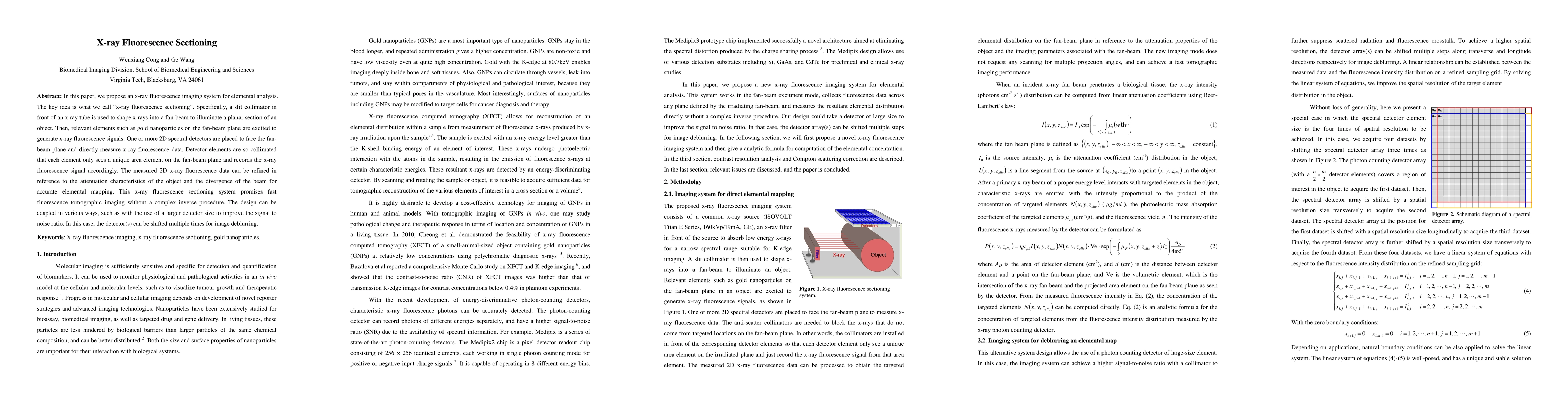
Specifically, a slit collimator in front of an x-ray tube is used to shape x-rays into a fan-beam to illuminate a planar section of an object. Then, relevant elements such as gold nanoparticles on the fan-beam plane are excited to generate x-ray fluorescence signals. One or more 2D spectral detectors are placed to face the fan-beam plane and directly measure x-ray fluorescence data. Detector elements are so collimated that each element only sees a unique area element on the fan-beam plane and records the x-ray fluorescence signal accordingly. The measured 2D x-ray fluorescence data can be refined in reference to the attenuation characteristics of the object and the divergence of the beam for accurate elemental mapping. This x-ray fluorescence sectioning system promises fast fluorescence tomographic imaging without a complex inverse procedure. The design can be adapted in various ways, such as with the use of a larger detector size to improve the signal to noise ratio. In this case, the detector(s) can be shifted multiple times for image deblurring.

AI Key Findings

Get AI-generated insights about this paper's methodology, results, and significance.

Paper Details

PDF Preview

Download PDF

Key Terms

fluorescence (0.403)
beam (0.263)
ray (0.256)
detector (0.211)
plane (0.183)
element (0.171)
imaging (0.159)
object (0.156)
elements (0.153)
signal (0.149)
records (0.131)
gold (0.131)
tube (0.131)
nanoparticles (0.130)
noise ratio (0.124)
refined (0.122)
placed (0.121)
divergence (0.121)
signal noise (0.120)
adapted (0.120)

Citation Network

Current paper (gray), citations (green), references (blue)

Current Paper
Citations
References
Click to view
Title Authors Year Actions
Development and characterisation of a confocal detection array for K-lines of heavy metals in big light matrix
J. Guarda, M. Valente, Rodolfo Figueroa
2021
View Paper
Quantitative Imaging of Gd Nanoparticles in Mice Using Benchtop Cone-Beam X-ray Fluorescence Computed Tomography System
Siyuan Zhang, Liang Li, Jiayou Chen, Zhiqiang Chen, Wenli Zhang, Hongbing Lu
2019
View Paper
Characteristic performance investigation of a photon counting detector for x-ray fluorescence imaging applications
L. Ren, Muhammad U. Ghani, Di Wu, Yuhua Li, Wei R. Chen, Ge Wang, B. Zheng, Hong Liu
2017
View Paper
Three-dimensional x-ray fluorescence mapping of a gold nanoparticle-loaded phantom.
L. Ren, Di Wu, Yuhua Li, Ge Wang, Xizeng Wu, Hong Liu
2014
View Paper

Comments (0)

This website uses cookies to ensure you get the best experience. Learn more