X-ray Free Electron Laser based Dark-Field X-ray Microscopy
Publication
Metrics
AI Quick Summary
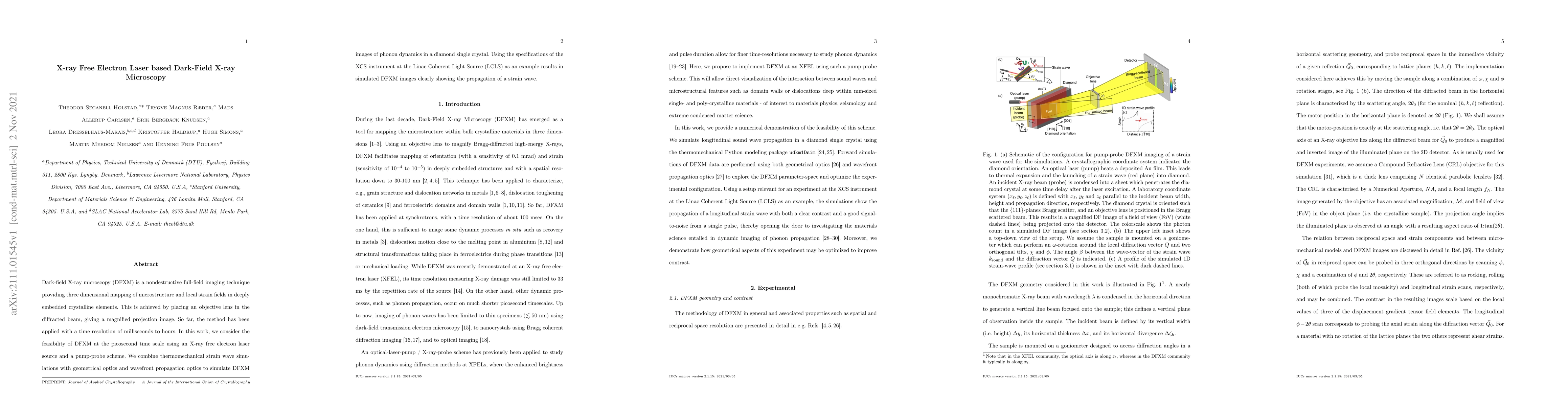
This paper explores the potential of X-ray free electron laser-based dark-field X-ray microscopy (DFXM) for capturing picosecond-timescale phonon dynamics. Simulations combining DFXM with thermomechanical strain wave models and optics demonstrate the method's capability to visualize strain wave propagation in a diamond crystal.
Paper Preview
Abstract
Dark-field X-ray microscopy (DFXM) is a nondestructive full-field imaging technique providing three dimensional mapping of microstructure and local strain fields in deeply embedded crystalline elements. This is achieved by placing an objective lens in the diffracted beam, giving a magnified projection image. So far, the method has been applied with a time resolution of milliseconds to hours. In this work, we consider the feasibility of DFXM at the picosecond time scale using an X-ray free electron laser source and a pump-probe scheme. We combine thermomechanical strain wave simulations with geometrical optics and wavefront propagation optics to simulate DFXM images of phonon dynamics in a diamond single crystal. Using the specifications of the XCS instrument at the Linac Coherent Light Source (LCLS) as an example results in simulated DFXM images clearly showing the propagation of a strain wave.
AI Key Findings
Get AI-generated insights about this paper's methodology, results, significance, and more — seven facets brought into focus.
Impact
Paper Details
Authors
PDF Preview
Key Terms
Citation Network
Current paper (gray), citations (green), references (blue)
Display is limited for performance on very large graphs.
Discussion 0