Publication
Metrics
AI Quick Summary
This paper demonstrates the use of synchrotron X-ray holographic tomography to non-destructively analyze 3D silicon photonic band gap crystals, revealing their internal 3D density distributions with 20 nanometer resolution. The study highlights how X-ray imaging can identify the functional discrepancies among similar-looking structures, underscoring its importance in assessing the quality of 3D photonic nanostructures.
Paper Preview
Abstract
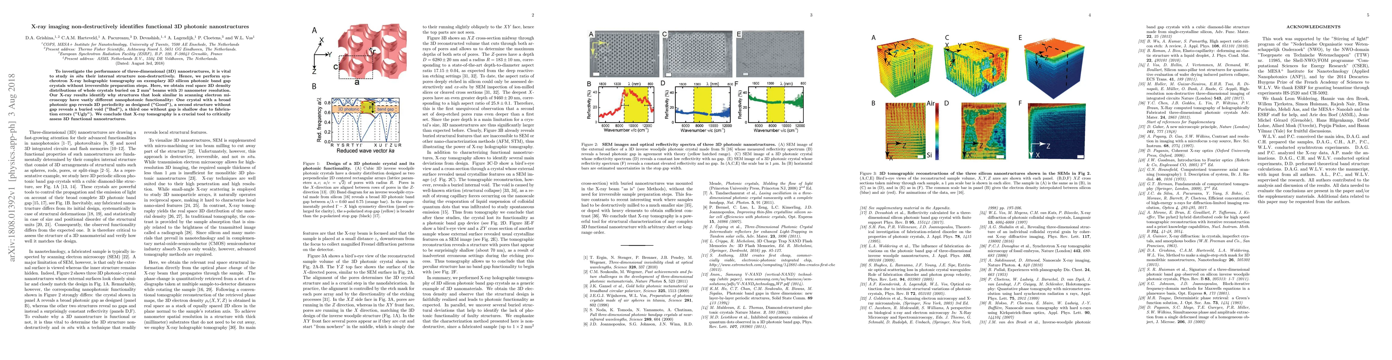
To investigate the performance of three-dimensional (3D) nanostructures, it is vital to study in situ their internal structure non-destructively. Hence, we perform synchrotron X-ray holographic tomography on exemplary 3D silicon photonic band gap crystals without irreversible preparation steps. Here, we obtain real space 3D density distributions of whole crystals buried on 2 mm^2 beams with 20 nanometer resolution. Our X-ray results identify why structures that look similar in scanning electron microscopy have vastly different nanophotonic functionality: One crystal with a broad photonic gap reveals 3D periodicity as designed ("Good"), a second structure without gap reveals a buried void ("Bad"), a third one without gap is shallow due to fabrication errors ("Ugly"). We conclude that X-ray tomography is a crucial tool to critically assess 3D functional nanostructures.
AI Key Findings
Get AI-generated insights about this paper's methodology, results, significance, and more — seven facets brought into focus.
Impact
Paper Details
Authors
PDF Preview
Key Terms
Citation Network
Current paper (gray), citations (green), references (blue)
Display is limited for performance on very large graphs.
Discussion 0