Publication
Metrics
Paper Preview
Abstract
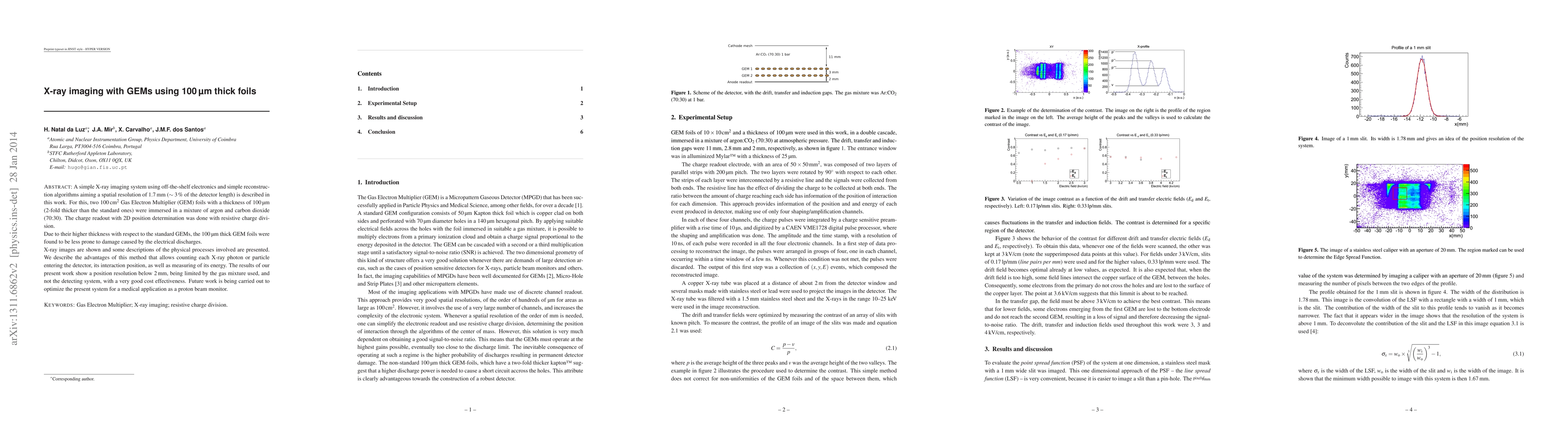
A simple X-ray imaging system using off-the-shelf electronics and simple reconstruction algorithms aiming a spatial resolution of 1.7 mm ($\sim 3\,\%$ of the detector length) is described in this work. For this, two 100 cm$^2$ Gas Electron Multiplier (GEM) foils with a thickness of 100 \mu m (2-fold thicker than the standard ones) were immersed in a mixture of argon and carbon dioxide (70:30). The charge readout with 2D position determination was done with resistive charge division. Due to their higher thickness with respect to the standard GEMs, the 100 \mu m thick GEM foils were found to be less prone to damage caused by the electrical discharges. X-ray images are shown and some descriptions of the physical processes involved are presented. We describe the advantages of this method that allows counting each X-ray photon or particle entering the detector, its interaction position, as well as measuring of its energy. The results of our present work show a position resolution below 2 mm, being limited by the gas mixture used, and not the detecting system, with a very good cost effectiveness. Future work is being carried out to optimize the present system for a medical application as a proton beam monitor.
AI Key Findings
Get AI-generated insights about this paper's methodology, results, significance, and more — seven facets brought into focus.
Impact
Paper Details
PDF Preview
Key Terms
Citation Network
Current paper (gray), citations (green), references (blue)
Display is limited for performance on very large graphs.
Discussion 0