X-ray projection imaging of metal oxide particles inside gingival tissues
Publication
Metrics
AI Quick Summary
This paper proposes using multiple energy X-ray projection imaging to detect and differentiate metal oxide particles, such as silicon dioxide and titanium dioxide, embedded in gingival tissues, which can cause foreign body gingivitis. Simulation results using GATE software indicate the feasibility of detecting particles as small as 0.5 micrometers with optimized imaging parameters.
Paper Preview
Abstract
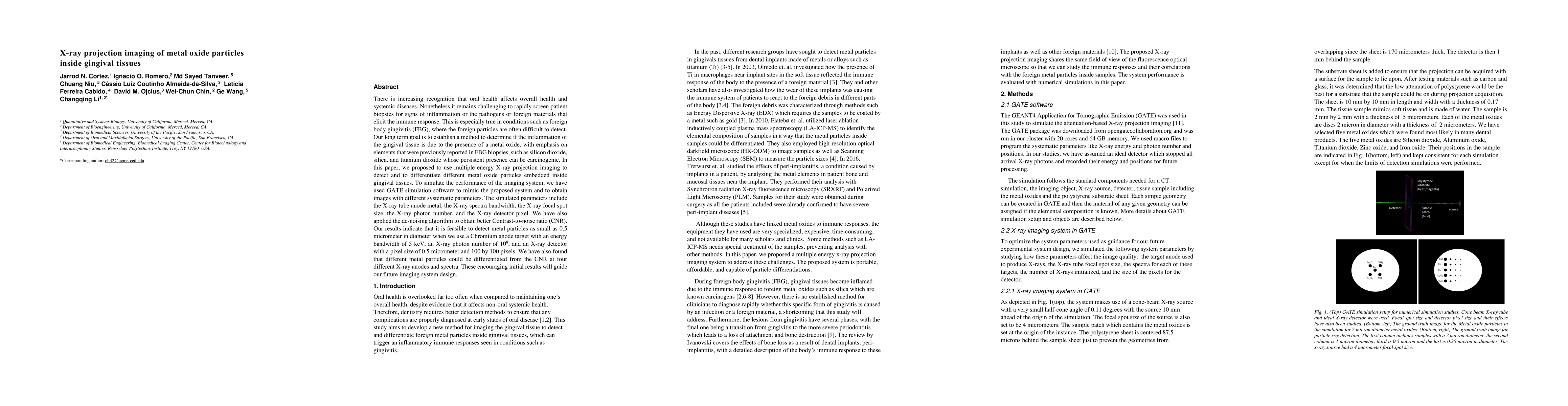
There is increasing recognition that oral health affects overall health and systemic diseases. Nonetheless it remains challenging to rapidly screen patient biopsies for signs of inflammation or the pathogens or foreign materials that elicit the immune response. This is especially true in conditions such as foreign body gingivitis (FBG), where the foreign particles are often difficult to detect. Our long term goal is to establish a method to determine if the inflammation of the gingival tissue is due to the presence of a metal oxide, with emphasis on elements that were previously reported in FBG biopsies, such as silicon dioxide, silica, and titanium dioxide whose persistent presence can be carcinogenic. In this paper, we proposed to use multiple energy X-ray projection imaging to detect and to differentiate different metal oxide particles embedded inside gingival tissues. To simulate the performance of the imaging system, we have used GATE simulation software to mimic the proposed system and to obtain images with different systematic parameters. The simulated parameters include the X-ray tube anode metal, the X-ray spectra bandwidth, the X-ray focal spot size, the X-ray photon number, and the X-ray dector pixel. We have also applied the de-noising algorithm to obtain better Contrast-to-noise ratio (CNR). Our results indicate that it is feasible to detect metal particles as small as 0.5 micrometer in diameter when we use a Chromium anode target with an energy bandwidth of 5 keV, an X-ray photon number of 10^8, and an X-ray detector with a pixel size of 0.5 micrometer and 100 by 100 pixels. We have also found that different metal particles could be differentiated from the CNR at four different X-ray anodes and spectra. These encouraging initial results will guide our future imaging system design.
AI Key Findings
Get AI-generated insights about this paper's methodology, results, significance, and more — seven facets brought into focus.
Impact
Paper Details
Authors
PDF Preview
Key Terms
Citation Network
Current paper (gray), citations (green), references (blue)
Display is limited for performance on very large graphs.
Discussion 0