X-Ray sum frequency generation; direct imaging of ultrafast electron dynamics
Publication
Metrics
AI Quick Summary
Researchers developed a new X-ray technique that captures ultrafast changes in electron density after optical excitation, revealing insights into molecular dynamics. This method uses a novel free electron laser source to produce spatial images of valence electron excitations.
Paper Preview
Abstract
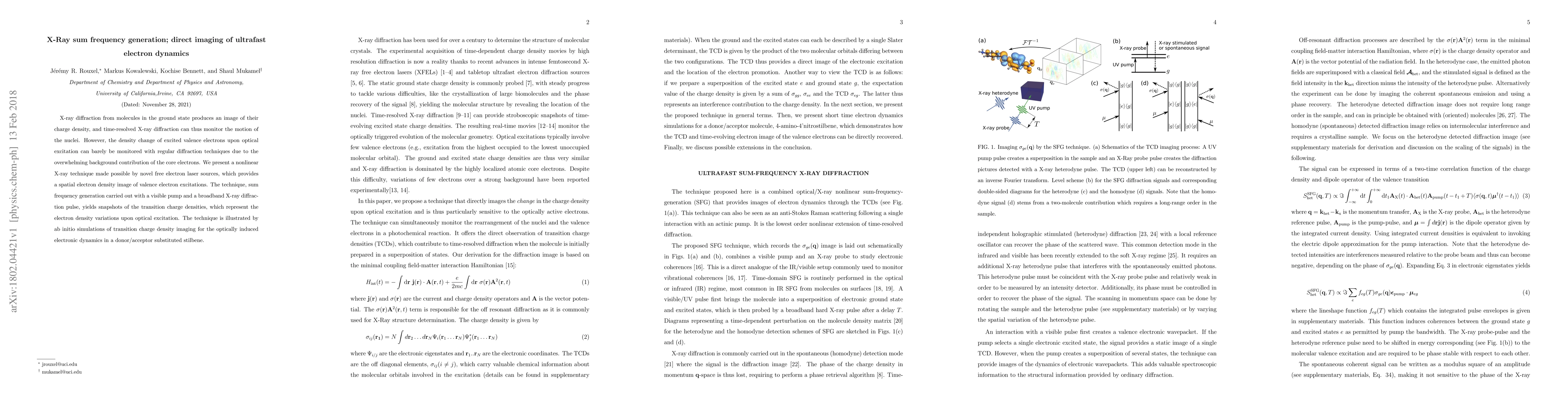
X-ray diffraction from molecules in the ground state produces an image of their charge density, and time-resolved X-ray diffraction can thus monitor the motion of the nuclei. However, the density change of excited valence electrons upon optical excitation can barely be monitored with regular diffraction techniques due to the overwhelming background contribution of the core electrons. We present a nonlinear X-ray technique made possible by novel free electron laser sources, which provides a spatial electron density image of valence electron excitations. The technique, sum frequency generation carried out with a visible pump and a broadband X-ray diffraction pulse, yields snapshots of the transition charge densities, which represent the electron density variations upon optical excitation. The technique is illustrated by ab initio simulations of transition charge density imaging for the optically induced electronic dynamics in a donor/acceptor substituted stilbene.
AI Key Findings
Get AI-generated insights about this paper's methodology, results, significance, and more — seven facets brought into focus.
Impact
Paper Details
PDF Preview
Key Terms
Citation Network
Current paper (gray), citations (green), references (blue)
Display is limited for performance on very large graphs.
Discussion 0