Background: Precise breast ultrasound (BUS) segmentation supports reliable
measurement, quantitative analysis, and downstream classification, yet remains
difficult for small or low-contrast lesions with fuzzy margins and speckle
noise. Text prompts can add clinical context, but directly applying weakly
localized text-image cues (e.g., CAM/CLIP-derived signals) tends to produce
coarse, blob-like responses that smear boundaries unless additional mechanisms
recover fine edges. Methods: We propose XBusNet, a novel dual-prompt,
dual-branch multimodal model that combines image features with clinically
grounded text. A global pathway based on a CLIP Vision Transformer encodes
whole-image semantics conditioned on lesion size and location, while a local
U-Net pathway emphasizes precise boundaries and is modulated by prompts that
describe shape, margin, and Breast Imaging Reporting and Data System (BI-RADS)
terms. Prompts are assembled automatically from structured metadata, requiring
no manual clicks. We evaluate on the Breast Lesions USG (BLU) dataset using
five-fold cross-validation. Primary metrics are Dice and Intersection over
Union (IoU); we also conduct size-stratified analyses and ablations to assess
the roles of the global and local paths and the text-driven modulation.
Results: XBusNet achieves state-of-the-art performance on BLU, with mean Dice
of 0.8765 and IoU of 0.8149, outperforming six strong baselines. Small lesions
show the largest gains, with fewer missed regions and fewer spurious
activations. Ablation studies show complementary contributions of global
context, local boundary modeling, and prompt-based modulation. Conclusions: A
dual-prompt, dual-branch multimodal design that merges global semantics with
local precision yields accurate BUS segmentation masks and improves robustness
for small, low-contrast lesions.
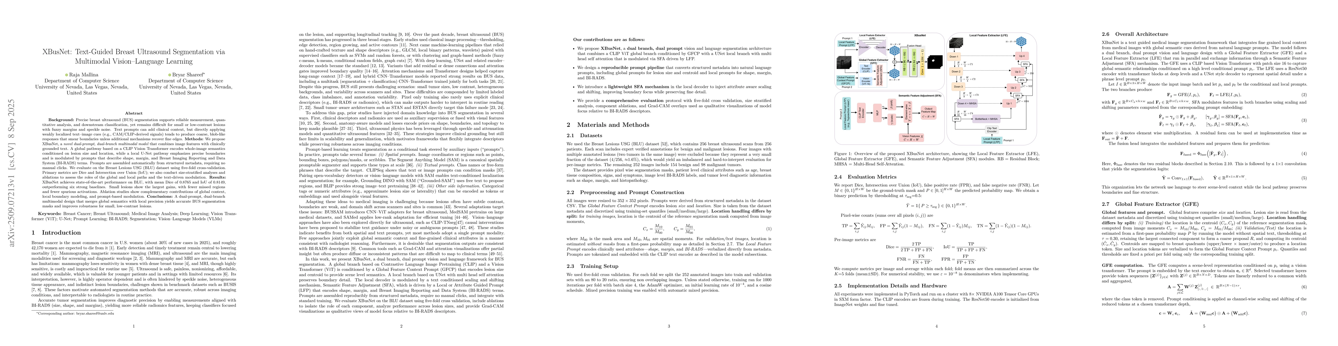
Discussion 0