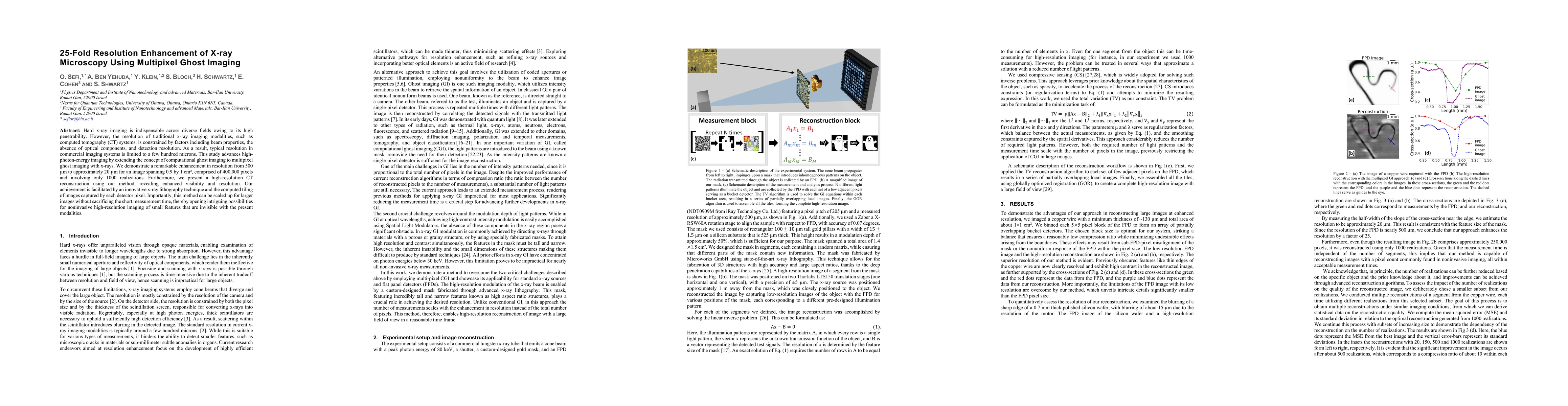
01
MethodologyHow they did it
Brief description of the research methodology used
This study enhances x-ray microscopy resolution by 25-fold using multipixel ghost imaging, achieving a resolution of approximately 20 microns from 500 microns. The method employs computational ghost imaging, innovative x-ray lithography, and tiled image reconstruction, enabling high-resolution CT scans and potential for noninvasive imaging of small features.
This study enhances x-ray microscopy resolution by 25-fold using multipixel ghost imaging, achieving a resolution of approximately 20 microns from 500 microns. The method employs computational ghost imaging, innovative x-ray lithography, and tiled image reconstruction, enabling high-resolution CT scans and potential for noninvasive imaging of small features.
Brief description of the research methodology used More in Methodology →
Main finding 1 — Main finding 2 More in Key Results →
Why this research is important and its potential impact More in Significance →
Limitation 1 — Limitation 2 More in Limitations →
Hard x-ray imaging is indispensable across diverse fields owing to its high penetrability. However, the resolution of traditional x-ray imaging modalities, such as computed tomography (CT) systems, is constrained by factors including beam properties, the absence of optical components, and detection resolution. As a result, typical resolution in commercial imaging systems is limited to a few hundred microns. This study advances high-photon-energy imaging by extending the concept of computational ghost imaging to multipixel ghost imaging with x-rays. We demonstrate a remarkable enhancement in resolution from 500 microns to approximately 20 microns for an image spanning 0.9 by 1 cm^2, comprised of 400,000 pixels and involving only 1000 realizations. Furthermore, we present a high-resolution CT reconstruction using our method, revealing enhanced visibility and resolution. Our achievement is facilitated by an innovative x-ray lithography technique and the computed tiling of images captured by each detector pixel. Importantly, this method can be scaled up for larger images without sacrificing the short measurement time, thereby opening intriguing possibilities for noninvasive high-resolution imaging of small features that are invisible with the present modalities.
Seven facets of this paper, analysed and brought into focus by AI.
Why this research is important and its potential impact
Brief description of the research methodology used
Why this research is important and its potential impact
Main technical or theoretical contribution
What makes this work novel or different from existing research
Current paper (gray), citations (green), references (blue)
Display is limited for performance on very large graphs.
Discussion 0