3D diffractive imaging of nanoparticle ensembles using an X-ray laser
Publication
Metrics
AI Quick Summary
This paper details the 3D structure determination of gold nanoparticles using X-ray single particle imaging with an X-ray laser, achieving unprecedented data volume and resolution of better than 3 nm. The study leverages the European X-ray free electron laser's capabilities to capture 1500 frames per second, enabling significant advancements in nanoparticle imaging techniques.
Paper Preview
Abstract
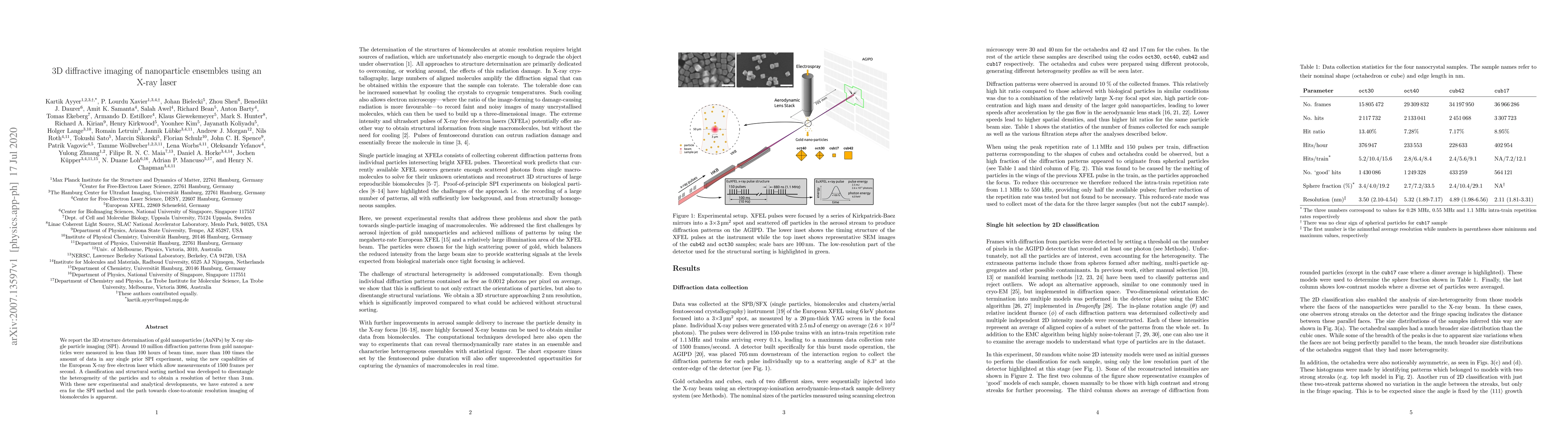
We report the 3D structure determination of gold nanoparticles (AuNPs) by X-ray single particle imaging (SPI). Around 10 million diffraction patterns from gold nanoparticles were measured in less than 100 hours of beam time, more than 100 times the amount of data in any single prior SPI experiment, using the new capabilities of the European X-ray free electron laser which allow measurements of 1500 frames per second. A classification and structural sorting method was developed to disentangle the heterogeneity of the particles and to obtain a resolution of better than 3 nm. With these new experimental and analytical developments, we have entered a new era for the SPI method and the path towards close-to-atomic resolution imaging of biomolecules is apparent.
AI Key Findings
Get AI-generated insights about this paper's methodology, results, significance, and more — seven facets brought into focus.
Impact
Paper Details
Authors
PDF Preview
Key Terms
Citation Network
Current paper (gray), citations (green), references (blue)
Display is limited for performance on very large graphs.
Discussion 0