A Deep Attentive Convolutional Neural Network for Automatic Cortical Plate Segmentation in Fetal MRI
Publication
Metrics
AI Quick Summary
This paper presents a deep attentive convolutional neural network for automatic segmentation of the fetal cortical plate in MRI scans, addressing the challenges of low resolution and morphological variations. The proposed method, utilizing deep supervision and residual connections, outperforms existing models and achieves high accuracy, facilitating large-scale studies on fetal brain development.
Paper Preview
Abstract
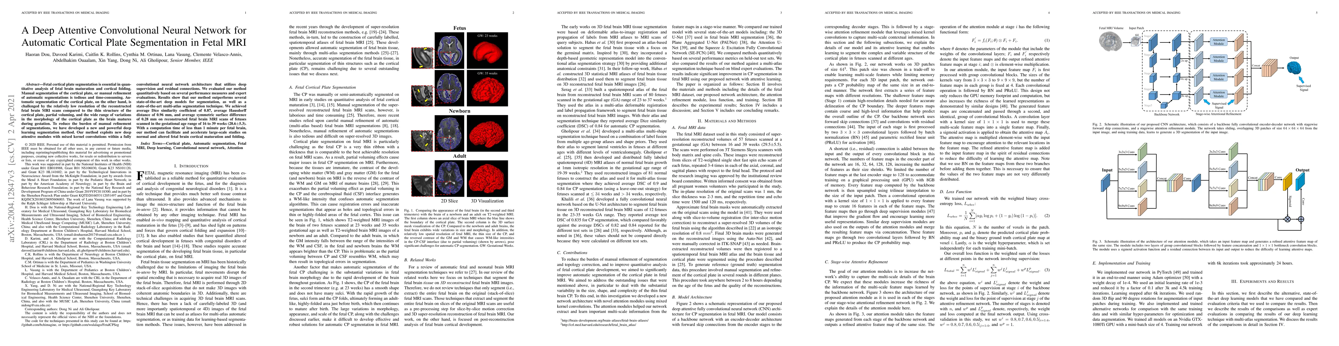
Fetal cortical plate segmentation is essential in quantitative analysis of fetal brain maturation and cortical folding. Manual segmentation of the cortical plate, or manual refinement of automatic segmentations is tedious and time-consuming. Automatic segmentation of the cortical plate, on the other hand, is challenged by the relatively low resolution of the reconstructed fetal brain MRI scans compared to the thin structure of the cortical plate, partial voluming, and the wide range of variations in the morphology of the cortical plate as the brain matures during gestation. To reduce the burden of manual refinement of segmentations, we have developed a new and powerful deep learning segmentation method. Our method exploits new deep attentive modules with mixed kernel convolutions within a fully convolutional neural network architecture that utilizes deep supervision and residual connections. We evaluated our method quantitatively based on several performance measures and expert evaluations. Results show that our method outperforms several state-of-the-art deep models for segmentation, as well as a state-of-the-art multi-atlas segmentation technique. We achieved average Dice similarity coefficient of 0.87, average Hausdorff distance of 0.96 mm, and average symmetric surface difference of 0.28 mm on reconstructed fetal brain MRI scans of fetuses scanned in the gestational age range of 16 to 39 weeks. With a computation time of less than 1 minute per fetal brain, our method can facilitate and accelerate large-scale studies on normal and altered fetal brain cortical maturation and folding.
AI Key Findings
Get AI-generated insights about this paper's methodology, results, significance, and more — seven facets brought into focus.
Impact
Paper Details
PDF Preview
Key Terms
Citation Network
Current paper (gray), citations (green), references (blue)
Display is limited for performance on very large graphs.
Discussion 0