ArxivLens

Publication

Published: Mar 28, 2025
Categories: eess.IV, cs.CV, 68T07

Metrics

Source: ArXiv

AI Quick Summary

This study developed an AI-driven system for detecting and grading osteoarthritis from knee X-rays using 1.3 million images from diverse Indian hospitals. The AI system showed high diagnostic accuracy and generalizability, promising to improve bone health diagnostics in resource-limited settings.

Quick Answers

What is "A Multi-Site Study on AI-Driven Pathology Detection and Osteoarthritis Grading from Knee X-Ray" about?

This study developed an AI-driven system for detecting and grading osteoarthritis from knee X-rays using 1.3 million images from diverse Indian hospitals. The AI system showed high diagnostic accuracy and generalizability, promising to improve bone health diagnostics in resource-limited settings.

What methodology did the authors use?

This study employed a multi-site clinical trial across various healthcare facilities in India, utilizing a dataset of over 51,000 knee X-ray scans. The dataset encompassed a wide range of imaging conditions, from high-resolution scans in advanced private hospitals to lower-quality images typical of government and SME facilities. The AI system was trained with tailored models for detecting specific pathologies and... More in Methodology →

What are the key results?

The AI-powered system demonstrated robust performance across diverse imaging conditions, equipment variability, and demographic subgroups. — Pathology-specific models achieved strong performance metrics for detecting joint space narrowing, sclerosis, osteophytes, alignment issues, soft tissue anomalies, and tibial spikes. More in Key Results →

Why is this work significant?

This research presents a scalable, cost-effective solution for bone health diagnostics, which holds promise for widespread adoption, especially in resource-limited healthcare settings. It transforms bone health management and enables proactive patient care by automating the detection and grading process, improving diagnostic efficiency, and reducing reliance on manual evaluations. More in Significance →

What are the main limitations?

The dataset, although diverse, lacks validation across international cohorts, which could enhance the model's generalizability. — Further exploration is needed to assess the system's integration into clinical workflows and its impact on treatment decisions. More in Limitations →

Paper Preview

Abstract

Introduction: Bone health disorders like osteoarthritis and osteoporosis pose major global health challenges, often leading to delayed diagnoses due to limited diagnostic tools. This study presents an AI-powered system that analyzes knee X-rays to detect key pathologies, including joint space narrowing, sclerosis, osteophytes, tibial spikes, alignment issues, and soft tissue anomalies. It also grades osteoarthritis severity, enabling timely, personalized treatment. 
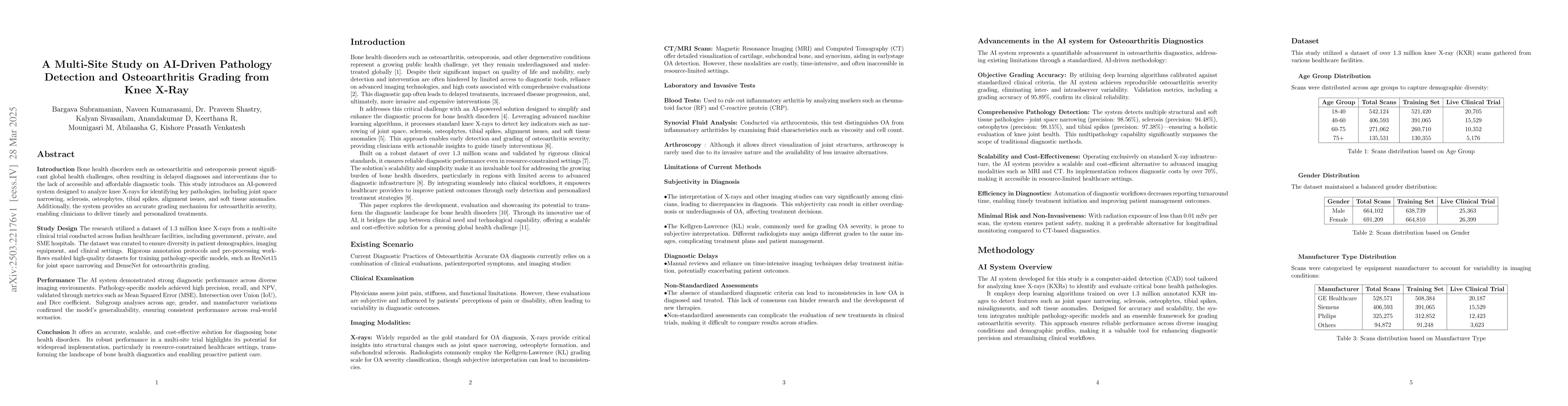
Study Design: The research used 1.3 million knee X-rays from a multi-site Indian clinical trial across government, private, and SME hospitals. The dataset ensured diversity in demographics, imaging equipment, and clinical settings. Rigorous annotation and preprocessing yielded high-quality training datasets for pathology-specific models like ResNet15 for joint space narrowing and DenseNet for osteoarthritis grading. Performance: The AI system achieved strong diagnostic accuracy across diverse imaging environments. Pathology-specific models excelled in precision, recall, and NPV, validated using Mean Squared Error (MSE), Intersection over Union (IoU), and Dice coefficient. Subgroup analyses across age, gender, and manufacturer variations confirmed generalizability for real-world applications. Conclusion: This scalable, cost-effective solution for bone health diagnostics demonstrated robust performance in a multi-site trial. It holds promise for widespread adoption, especially in resource-limited healthcare settings, transforming bone health management and enabling proactive patient care.

Key Findings, in focus

Seven facets of this paper, analysed and brought into focus by AI.

AI-generated · verify against source Generated Jun 10, 2025
In focus · one-line synthesis

This research presents a scalable, cost-effective solution for bone health diagnostics, which holds promise for widespread adoption, especially in resource-limited healthcare settings.

01
MethodologyHow they did it

This study employed a multi-site clinical trial across various healthcare facilities in India, utilizing a dataset of over 51,000 knee X-ray scans. The dataset encompassed a wide range of imaging conditions, from high-resolution scans in advanced private hospitals to lower-quality images typical of government and SME facilities. The AI system was trained with tailored models for detecting specific pathologies and grading osteoarthritis severity, validated through precision, recall, and NPV.

02
ResultsWhat they found
  • The AI-powered system demonstrated robust performance across diverse imaging conditions, equipment variability, and demographic subgroups.
  • Pathology-specific models achieved strong performance metrics for detecting joint space narrowing, sclerosis, osteophytes, alignment issues, soft tissue anomalies, and tibial spikes.
  • Subgroup analysis confirmed consistent accuracy, precision, and recall across age, gender, and manufacturer variations.
03
SignificanceWhy it matters

This research presents a scalable, cost-effective solution for bone health diagnostics, which holds promise for widespread adoption, especially in resource-limited healthcare settings. It transforms bone health management and enables proactive patient care by automating the detection and grading process, improving diagnostic efficiency, and reducing reliance on manual evaluations.

04
Technical ContributionsWhat's new technically

The development and validation of an AI-powered system for knee X-ray analysis, designed to detect critical pathologies and grade osteoarthritis severity with high accuracy.

05
NoveltyWhat sets it apart

This work stands out by addressing long-standing challenges in traditional workflows, such as variability in radiological interpretations and delays in diagnosis, through automated detection and grading processes.

06
LimitationsWhat to be cautious about
  • The dataset, although diverse, lacks validation across international cohorts, which could enhance the model's generalizability.
  • Further exploration is needed to assess the system's integration into clinical workflows and its impact on treatment decisions.
07
Future WorkWhere it goes next
  • Broader clinical validation and workflow integration should be pursued to fully realize the system's transformative impact on early diagnosis and improved patient care.
  • Investigating the system's performance in diverse international cohorts could further strengthen its generalizability.

Impact

20
References

Paper Details

Paper ID: 2503.22176
Comments: 15 pages, 2 figures
Categories:
eess.IV cs.CV 68T07

How to Cite This Paper

Reference manager: RIS EndNote

Citation Network

Current paper (gray), citations (green), references (blue)

Display is limited for performance on very large graphs.

Current Paper
Citations
References
Click to view


Discussion 0

This website uses cookies to ensure you get the best experience. Learn more