ArxivLens

Publication

Published: Mar 22, 2025
Categories: eess.IV

Metrics

Source: ArXiv

AI Quick Summary

This paper proposes a deep learning-refined subspace model for accelerated cardiac parametric mapping, improving spatial basis estimation and image fidelity. It combines low-rank tensor models with deep neural networks to enhance the accuracy of cardiac fibrosis and edema evaluation, compared against traditional Fourier-based reconstructions.

Quick Answers

What methodology did the authors use?

The research proposes a deep learning-refined subspace model for accelerated cardiac parametric mapping, utilizing a low-rank tensor model and a deep neural network to refine spatial basis images and temporal basis time-courses, improving fidelity and reducing noise in undersampled reconstruction. More in Methodology →

What are the key results?

The proposed method outperformed conventional parallel imaging reconstruction in terms of NRMSE, PSNR, NMSE, and SSIM for T1 and T2 mapping. — The method successfully reduced residual blurring and aliasing artifacts, especially in T2-weighted images, even at high acceleration factors (R=10). More in Key Results →

Why is this work significant?

This research is significant as it introduces a novel framework combining parallel imaging, temporal low-rank constraints, and deep learning for spatial restoration, which can improve image quality in highly accelerated cardiac parametric mapping sequences, potentially facilitating greater use of advanced reconstruction methods in standard clinical practice. More in Significance →

What are the main limitations?

The method was trained on data from a single vendor and specific T1 and T2 mapping sequences, so its generalizability to unknown sequences or contrasts has not been rigorously evaluated. — T2 mapping uses a full rank of 3, which improves parametric accuracy but reduces temporal compressibility, leading to slightly lower image quality at higher acceleration factors compared to T1... More in Limitations →

Paper Preview

Abstract

Cardiac parametric mapping is useful for evaluating cardiac fibrosis and edema. Parametric mapping relies on single-shot heartbeat-by-heartbeat imaging, which is susceptible to intra-shot motion during the imaging window. However, reducing the imaging window requires undersampled reconstruction techniques to preserve image fidelity and spatial resolution. The proposed approach is based on a low-rank tensor model of the multi-dimensional data, which jointly estimates spatial basis images and temporal basis time-courses from an auxiliary parallel imaging reconstruction. The tensor-estimated spatial basis is then further refined using a deep neural network, trained in a fully supervised fashion, improving the fidelity of the spatial basis using learned representations of cardiac basis functions. This two-stage spatial basis estimation will be compared against Fourier-based reconstructions and parallel imaging alone to demonstrate the sharpening and denoising properties of the deep learning-based subspace analysis.
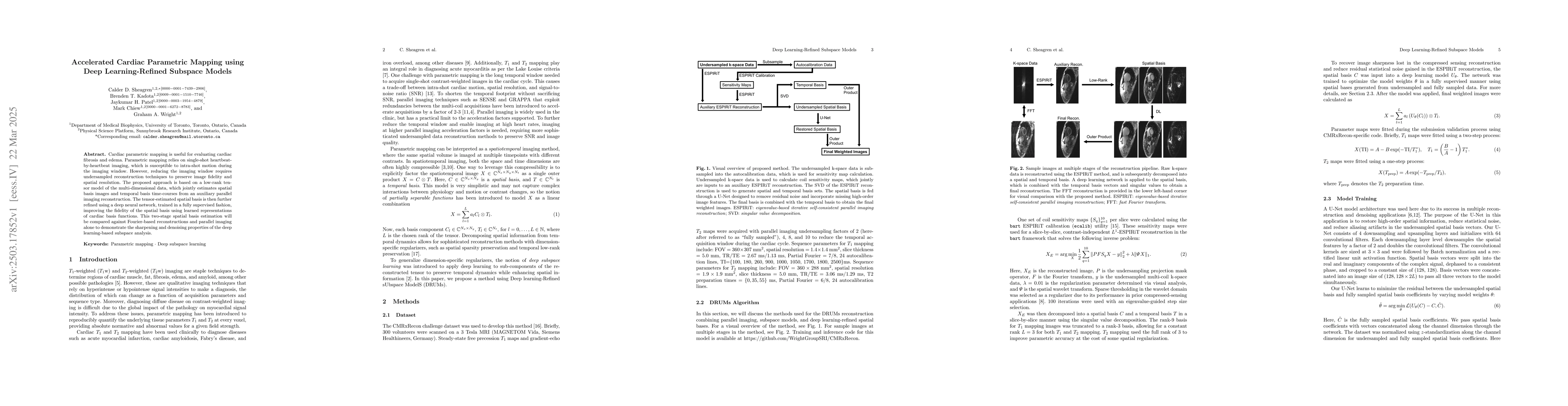
Key Findings, in focus

Seven facets of this paper, analysed and brought into focus by AI.

AI-generated · verify against source Generated Jun 10, 2025
In focus · one-line synthesis

This research is significant as it introduces a novel framework combining parallel imaging, temporal low-rank constraints, and deep learning for spatial restoration, which can improve image quality in highly accelerated cardiac parametric mapping sequences, potentially facilitating greater use of advanced reconstruction methods in standard clinical practice.

01
MethodologyHow they did it

The research proposes a deep learning-refined subspace model for accelerated cardiac parametric mapping, utilizing a low-rank tensor model and a deep neural network to refine spatial basis images and temporal basis time-courses, improving fidelity and reducing noise in undersampled reconstruction.

02
ResultsWhat they found
  • The proposed method outperformed conventional parallel imaging reconstruction in terms of NRMSE, PSNR, NMSE, and SSIM for T1 and T2 mapping.
  • The method successfully reduced residual blurring and aliasing artifacts, especially in T2-weighted images, even at high acceleration factors (R=10).
  • Quantitative results showed that the method generally performed better in T2 reconstruction than T1 reconstruction, with the exception of PSNR.
03
SignificanceWhy it matters

This research is significant as it introduces a novel framework combining parallel imaging, temporal low-rank constraints, and deep learning for spatial restoration, which can improve image quality in highly accelerated cardiac parametric mapping sequences, potentially facilitating greater use of advanced reconstruction methods in standard clinical practice.

04
Technical ContributionsWhat's new technically

The paper presents a deep learning-refined subspace model for cardiac parametric mapping, incorporating a low-rank tensor model and a deep neural network to refine spatial basis images and temporal basis time-courses, enhancing fidelity and reducing noise in undersampled reconstruction.

05
NoveltyWhat sets it apart

The novelty of this work lies in the integration of deep learning with a low-rank subspace model for spatial basis refinement, which improves image quality in accelerated cardiac parametric mapping sequences while enforcing temporal consistency through data-driven constraints.

06
LimitationsWhat to be cautious about
  • The method was trained on data from a single vendor and specific T1 and T2 mapping sequences, so its generalizability to unknown sequences or contrasts has not been rigorously evaluated.
  • T2 mapping uses a full rank of 3, which improves parametric accuracy but reduces temporal compressibility, leading to slightly lower image quality at higher acceleration factors compared to T1 mapping.
07
Future WorkWhere it goes next
  • Investigate the effect of rank-2 approximations for T2 multi-contrast images on overall image quality and parametric robustness.
  • Optimize the parallel imaging undersampling pattern for better temporal coherence of artifacts.

Impact

17
References

Paper Details

Paper ID: 2503.17852
Comments: 11 pages, 4 figures
Categories:
eess.IV

How to Cite This Paper

Reference manager: RIS EndNote

Citation Network

Current paper (gray), citations (green), references (blue)

Display is limited for performance on very large graphs.

Current Paper
Citations
References
Click to view


Discussion 0

This website uses cookies to ensure you get the best experience. Learn more