Publication
Metrics
AI Quick Summary
This paper introduces a cost-effective method for active illumination using a digital micromirror device (DMD) for quantitative phase imaging, generating plane waves with various angles for high-resolution phase imaging and refractive index tomography. The method is validated through imaging of colloidal spheres and biological cells.
Paper Preview
Abstract
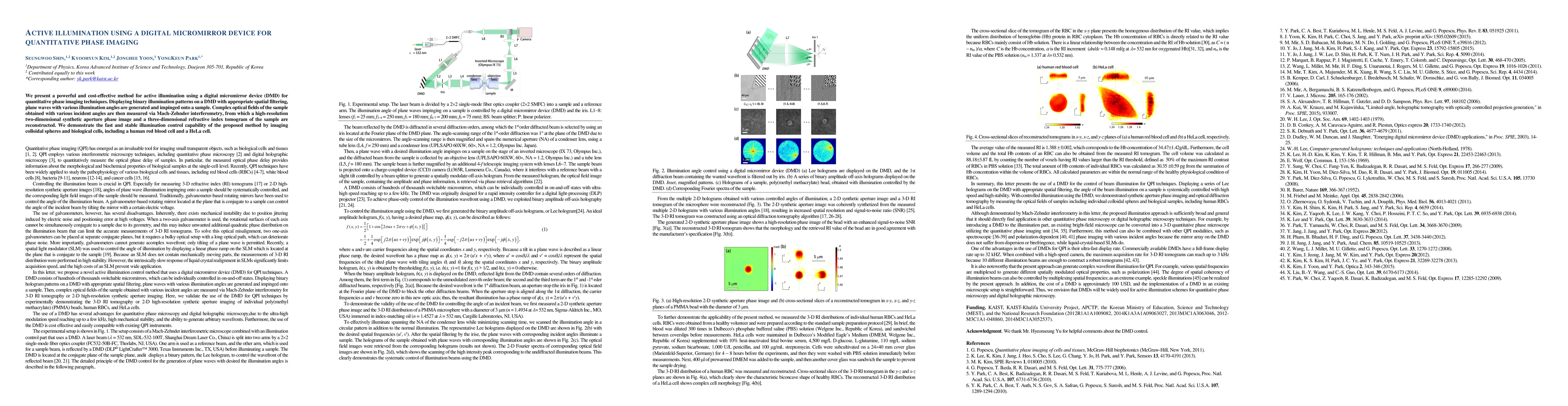
We present a powerful and cost-effective method for active illumination using a digital micromirror device (DMD) for quantitative phase imaging techniques. Displaying binary illumination patterns on a DMD with appropriate spatial filtering, plane waves with various illumination angles are generated and impinged onto a sample. Complex optical fields of the sample obtained with various incident angles are then measured via Mach-Zehnder interferometry, from which a high-resolution two-dimensional synthetic aperture phase image and a three-dimensional refractive index tomogram of the sample are reconstructed. We demonstrate the fast and stable illumination control capability of the proposed method by imaging colloidal spheres and biological cells, including a human red blood cell and a HeLa cell.
AI Key Findings
Get AI-generated insights about this paper's methodology, results, significance, and more — seven facets brought into focus.
Impact
Paper Details
Authors
PDF Preview
Key Terms
Citation Network
Current paper (gray), citations (green), references (blue)
Display is limited for performance on very large graphs.
Discussion 0