An Iterative Convolutional Neural Network Algorithm Improves Electron Microscopy Image Segmentation
Publication
Metrics
AI Quick Summary
Researchers developed an algorithm to improve electron microscopy image segmentation by iteratively refining a probability map of cell membranes, achieving significant improvements in membrane detection accuracy.
Paper Preview
Abstract
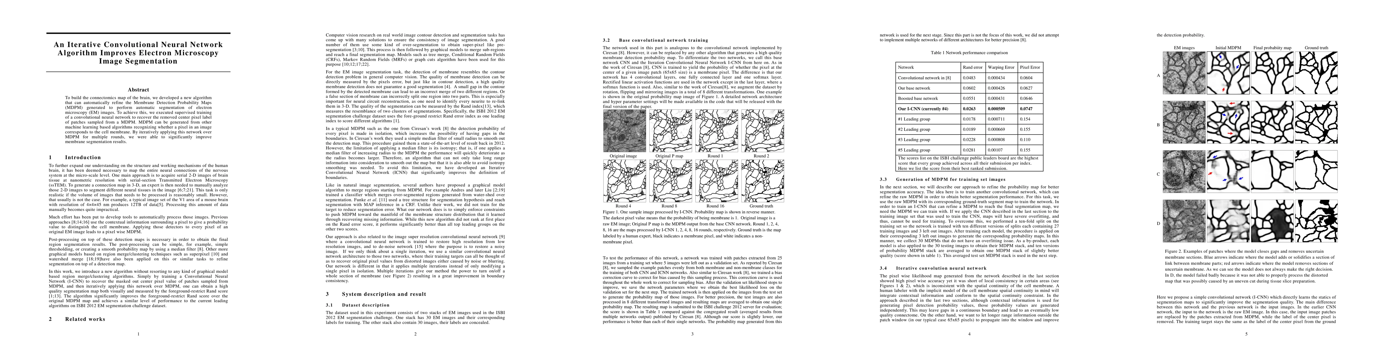
To build the connectomics map of the brain, we developed a new algorithm that can automatically refine the Membrane Detection Probability Maps (MDPM) generated to perform automatic segmentation of electron microscopy (EM) images. To achieve this, we executed supervised training of a convolutional neural network to recover the removed center pixel label of patches sampled from a MDPM. MDPM can be generated from other machine learning based algorithms recognizing whether a pixel in an image corresponds to the cell membrane. By iteratively applying this network over MDPM for multiple rounds, we were able to significantly improve membrane segmentation results.
AI Key Findings
Get AI-generated insights about this paper's methodology, results, significance, and more — seven facets brought into focus.
Impact
Paper Details
PDF Preview
Key Terms
Citation Network
Current paper (gray), citations (green), references (blue)
Display is limited for performance on very large graphs.
Discussion 0