Publication

Published: Oct 23, 2018
Updated: Jun 16, 2025
Categories: cs.CV

Metrics

Source: ArXiv

Quick Actions

View PDF

AI Quick Summary

This research presents a fully automated method for segmenting atrial scars in LGE MRI using a graph-cuts framework and Deep Neural Network (DNN). The method learned potentials from a surface mesh of the left atrium, improving segmentation accuracy with larger training patches. It outperformed published methods, demonstrating better results in quantifying scars, which could aid in atrial fibrillation diagnosis and prognosis.

Quick Answers

What is "Atrial scars segmentation via potential learning in the graph-cuts framework" about?

This research presents a fully automated method for segmenting atrial scars in LGE MRI using a graph-cuts framework and Deep Neural Network (DNN). The method learned potentials from a surface mesh of the left atrium, improving segmentation accuracy with larger training patches. It outperformed published methods, demonstrating better results in quantifying scars, which could aid in atrial fibrillation diagnosis and prognosis.

What methodology did the authors use?

The research proposes a fully automated method for atrial scars segmentation using a graph-cuts framework where potentials are learned via a Deep Neural Network (DNN) on a surface mesh of the left atrium (LA). The method utilizes equidistant projection and DNN for learning structural and texture information from patches. More in Methodology →

What are the key results?

The proposed method's performance improved and converged with increasing training patch sizes, indicating better feature learning. — Balancing the contribution from t-link and n-link improved segmentation results, thanks to the inter-relationship learned by the DNN for the graph-cuts algorithm. More in Key Results →

Why is this work significant?

This research is significant as it addresses the challenge of automating quantification and analysis of atrial scars in LGE MRI for atrial fibrillation (AF) patients, which can aid in diagnosis and prognosis of AF. More in Significance →

What are the main limitations?

The study was limited to 100 datasets with manual delineation for validation. — The method's performance may depend on the quality of the initial LA segmentation obtained from multi-atlas propagation and image registration. More in Limitations →

Paper Preview

Abstract

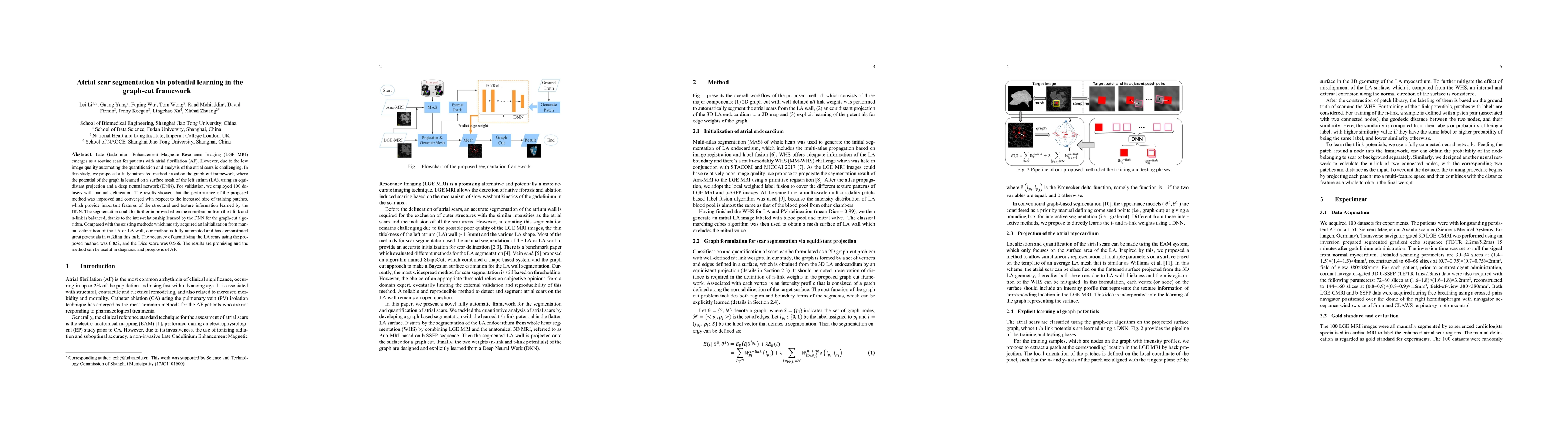
Late Gadolinium Enhancement Magnetic Resonance Imaging (LGE MRI) emerged as a routine scan for patients with atrial fibrillation (AF). However, due to the low image quality automating the quantification and analysis of the atrial scars is challenging. In this study, we pro-posed a fully automated method based on the graph-cuts framework, where the potential of the graph is learned on a surface mesh of the left atrium (LA) using an equidistant projection and a Deep Neural Network (DNN). For validation, we employed 100 datasets with manual delineation. The results showed that the performance of the proposed method improved and converged with respect to the increased size of training patches, which provide important features of the structural and texture information learned by the DNN. The segmentation could be further improved when the contribution from the t-link and n-link is balanced, thanks to inter-relationship learned by the DNN for the graph-cuts algorithm. Compared with the published methods which mostly acquired manual delineation of the LA or LA wall, our method is fully automatic and demonstrated evidently better results with statistical significance. Finally, the accuracy of quantifying the scars assessed by the Dice score was 0.570. The results are promising and the method can be useful in diagnosis and prognosis of AF.

AI Key Findings

Generated Sep 03, 2025

Methodology — What approach did the authors take?

The research proposes a fully automated method for atrial scars segmentation using a graph-cuts framework where potentials are learned via a Deep Neural Network (DNN) on a surface mesh of the left atrium (LA). The method utilizes equidistant projection and DNN for learning structural and texture information from patches.

Key Results — What are the main findings?

  • The proposed method's performance improved and converged with increasing training patch sizes, indicating better feature learning.
  • Balancing the contribution from t-link and n-link improved segmentation results, thanks to the inter-relationship learned by the DNN for the graph-cuts algorithm.
  • Compared to published methods, the proposed method demonstrated statistically significant better segmentation results with full automation.

Significance — Why does this research matter?

This research is significant as it addresses the challenge of automating quantification and analysis of atrial scars in LGE MRI for atrial fibrillation (AF) patients, which can aid in diagnosis and prognosis of AF.

Technical Contribution — What is the technical contribution?

The main technical contribution is the integration of a DNN to learn graph potentials in a graph-cuts framework for atrial scars segmentation, balancing t-link and n-link contributions for improved results.

Novelty — What is new about this work?

This work stands out by proposing a fully automated method for atrial scars segmentation, utilizing a DNN to learn graph potentials in a graph-cuts framework, outperforming existing published methods with statistical significance.

Limitations — What are the limitations of this study?

  • The study was limited to 100 datasets with manual delineation for validation.
  • The method's performance may depend on the quality of the initial LA segmentation obtained from multi-atlas propagation and image registration.

Future Work — What did the authors propose for future work?

  • Explore the method's applicability on larger and more diverse datasets.
  • Investigate the impact of different DNN architectures and training strategies on segmentation performance.

Paper Details

Paper ID: 1810.09123
License: http://arxiv.org/licenses/nonexclusive-distrib/1.0/
Comments: 9 pages,4 figures,STACOM2018
Categories:
cs.CV

How to Cite This Paper

BibTeX
@article{anon2018atrial,
  title         = {Atrial scars segmentation via potential learning in the graph-cuts
  framework},
  year          = {2018},
  eprint        = {1810.09123},
  archivePrefix = {arXiv},
  primaryClass  = {cs.CV},
}
APA 7
(2018). Atrial scars segmentation via potential learning in the graph-cuts
  framework. arXiv. https://arxiv.org/abs/1810.09123
MLA 9
"Atrial scars segmentation via potential learning in the graph-cuts
  framework." arXiv, 2018, arxiv.org/abs/1810.09123.

Key Terms

dnn (0.259)
la (0.254)
af (0.213)
cuts (0.211)
learned (0.207)
manual (0.195)
link (0.167)
graph (0.162)

Citation Network

Current paper (gray), citations (green), references (blue)

Display is limited for performance on very large graphs.

Current Paper
Citations
References
Click to view

No citations found for this paper.

Comments (0)

This website uses cookies to ensure you get the best experience. Learn more