Publication
Metrics
AI Quick Summary
Automated lung segmentation achieved similar results using different deep learning approaches, with a standard U-net method performing best on routine imaging data, suggesting that improving data diversity is key to increasing clinical applicability.
Paper Preview
Abstract
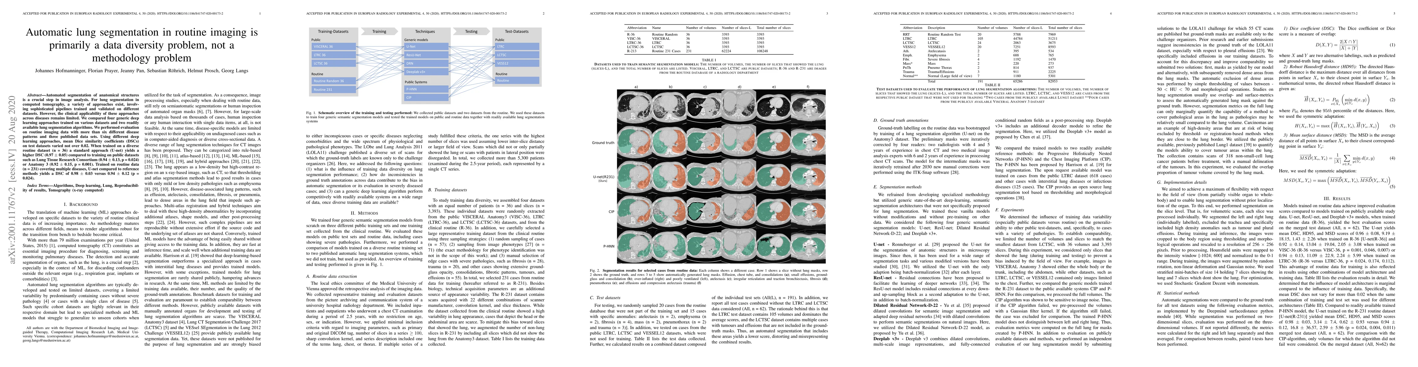
Automated segmentation of anatomical structures is a crucial step in image analysis. For lung segmentation in computed tomography, a variety of approaches exist, involving sophisticated pipelines trained and validated on different datasets. However, the clinical applicability of these approaches across diseases remains limited. We compared four generic deep learning approaches trained on various datasets and two readily available lung segmentation algorithms. We performed evaluation on routine imaging data with more than six different disease patterns and three published data sets. Using different deep learning approaches, mean Dice similarity coefficients (DSCs) on test datasets varied not over 0.02. When trained on a diverse routine dataset (n = 36) a standard approach (U-net) yields a higher DSC (0.97 $\pm$ 0.05) compared to training on public datasets such as Lung Tissue Research Consortium (0.94 $\pm$ 0.13, p = 0.024) or Anatomy 3 (0.92 $\pm$ 0.15, p = 0.001). Trained on routine data (n = 231) covering multiple diseases, U-net compared to reference methods yields a DSC of 0.98 $\pm$ 0.03 versus 0.94 $\pm$ 0.12 (p = 0.024).
AI Key Findings
Get AI-generated insights about this paper's methodology, results, significance, and more — seven facets brought into focus.
Impact
Paper Details
PDF Preview
Key Terms
Citation Network
Current paper (gray), citations (green), references (blue)
Display is limited for performance on very large graphs.
Discussion 0