Carotid artery wall segmentation in ultrasound image sequences using a deep convolutional neural network
Publication
Metrics
AI Quick Summary
This study proposes a deep convolutional neural network, specifically a dilated U-net, for automatic segmentation of the carotid artery wall in ultrasound images, achieving a mean absolute difference of <120 um compared to expert annotations. The method demonstrates a 98.7% success rate, suggesting its robustness and potential for clinical use.
Paper Preview
Abstract
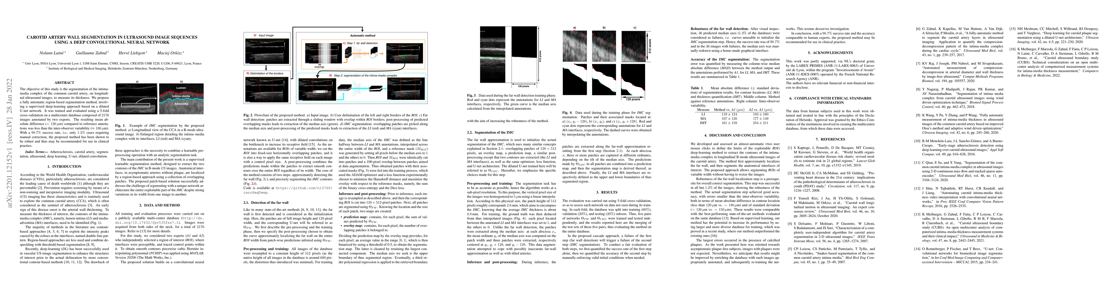
The objective of this study is the segmentation of the intima-media complex of the common carotid artery, on longitudinal ultrasound images, to measure its thickness. We propose a fully automatic region-based segmentation method, involving a supervised region-based deep-learning approach based on a dilated U-net network. It was trained and evaluated using a 5-fold cross-validation on a multicenter database composed of 2176 images annotated by two experts. The resulting mean absolute difference (<120 um) compared to reference annotations was less than the inter-observer variability (180 um). With a 98.7% success rate, i.e., only 1.3% cases requiring manual correction, the proposed method has been shown to be robust and thus may be recommended for use in clinical practice.
AI Key Findings
Get AI-generated insights about this paper's methodology, results, significance, and more — seven facets brought into focus.
Impact
Paper Details
Authors
PDF Preview
Key Terms
Citation Network
Current paper (gray), citations (green), references (blue)
Display is limited for performance on very large graphs.
Discussion 0