Combining unsupervised and supervised learning in microscopy enables defect analysis of a full 4H-SiC wafer
Publication
Metrics
AI Quick Summary
This paper combines unsupervised and supervised learning techniques to develop an automated image analysis pipeline for detecting and analyzing defects in a full 4H-SiC wafer using microscopy. The method enables accurate extraction of defect types and positions from a large, stitched image dataset.
Paper Preview
Abstract
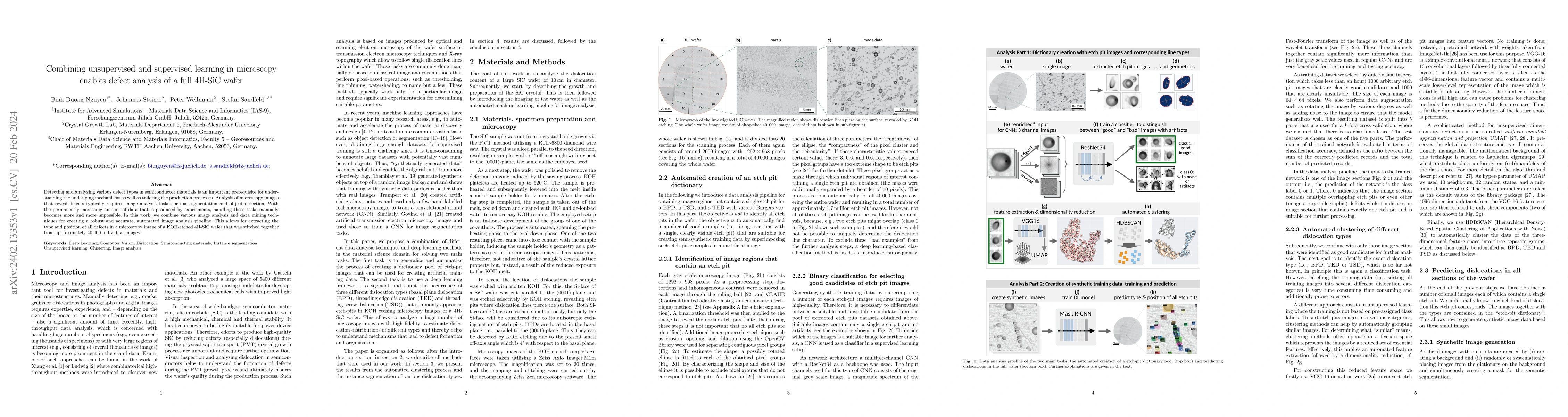
Detecting and analyzing various defect types in semiconductor materials is an important prerequisite for understanding the underlying mechanisms as well as tailoring the production processes. Analysis of microscopy images that reveal defects typically requires image analysis tasks such as segmentation and object detection. With the permanently increasing amount of data that is produced by experiments, handling these tasks manually becomes more and more impossible. In this work, we combine various image analysis and data mining techniques for creating a robust and accurate, automated image analysis pipeline. This allows for extracting the type and position of all defects in a microscopy image of a KOH-etched 4H-SiC wafer that was stitched together from approximately 40,000 individual images.
AI Key Findings
Get AI-generated insights about this paper's methodology, results, significance, and more — seven facets brought into focus.
Impact
Paper Details
Authors
PDF Preview
Key Terms
Citation Network
Current paper (gray), citations (green), references (blue)
Display is limited for performance on very large graphs.
Discussion 0