Compositional Representation Learning for Brain Tumour Segmentation
More tools
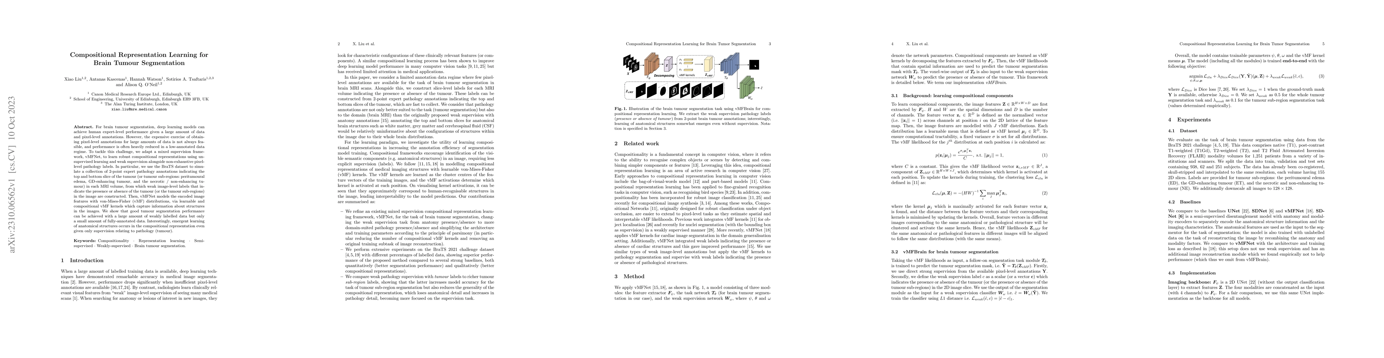
This paper proposes vMFNet, a mixed supervision framework for brain tumour segmentation that leverages unsupervised learning and weak supervision to achieve robust performance with minimal fully-annotated data. The method uses 2-point expert pathology annotations to generate weak image-level labels, enabling the learning of compositional representations via von-Mises-Fisher distributions, which captures structural information from the MRI images.
Abstract
For brain tumour segmentation, deep learning models can achieve human expert-level performance given a large amount of data and pixel-level annotations. However, the expensive exercise of obtaining pixel-level annotations for large amounts of data is not always feasible, and performance is often heavily reduced in a low-annotated data regime. To tackle this challenge, we adapt a mixed supervision framework, vMFNet, to learn robust compositional representations using unsupervised learning and weak supervision alongside non-exhaustive pixel-level pathology labels. In particular, we use the BraTS dataset to simulate a collection of 2-point expert pathology annotations indicating the top and bottom slice of the tumour (or tumour sub-regions: peritumoural edema, GD-enhancing tumour, and the necrotic / non-enhancing tumour) in each MRI volume, from which weak image-level labels that indicate the presence or absence of the tumour (or the tumour sub-regions) in the image are constructed. Then, vMFNet models the encoded image features with von-Mises-Fisher (vMF) distributions, via learnable and compositional vMF kernels which capture information about structures in the images. We show that good tumour segmentation performance can be achieved with a large amount of weakly labelled data but only a small amount of fully-annotated data. Interestingly, emergent learning of anatomical structures occurs in the compositional representation even given only supervision relating to pathology (tumour).
AI Key Findings
Get AI-generated insights about this paper's methodology, results, significance, and more — available when analysis has been generated.
Discussion 0