Authors
Publication
Metrics
Quick Actions
AI Quick Summary
A new method called DC-Seg is proposed to improve brain tumor segmentation with missing modalities by disentangling images into anatomical and modality-specific representations, outperforming state-of-the-art methods in experiments.
Quick Answers
What is "DC-Seg: Disentangled Contrastive Learning for Brain Tumor Segmentation with Missing Modalities" about?
A new method called DC-Seg is proposed to improve brain tumor segmentation with missing modalities by disentangling images into anatomical and modality-specific representations, outperforming state-of-the-art methods in experiments.
What methodology did the authors use?
The proposed method, DC-Seg, disentangles brain images into modality-invariant anatomical representation and modality-specific representation using anatomical contrastive learning and modality contrastive learning respectively. It also introduces a segmentation-based regularizer for handling missing modalities. More in Methodology →
What are the key results?
DC-Seg outperforms state-of-the-art methods in incomplete multimodal brain tumor segmentation tasks with varying missing modalities. — The method demonstrates strong generalizability in white matter hyperintensity (WMH) segmentation. More in Key Results →
Why is this work significant?
This research is important as it addresses the challenge of missing modalities in brain tumor segmentation, improving the accuracy and robustness of medical image analysis, which can lead to better clinical decision-making. More in Significance →
What are the main limitations?
The paper does not discuss potential limitations or challenges encountered during the research. — No information on scalability or computational resource requirements for the proposed method. More in Limitations →
Paper Preview
Abstract
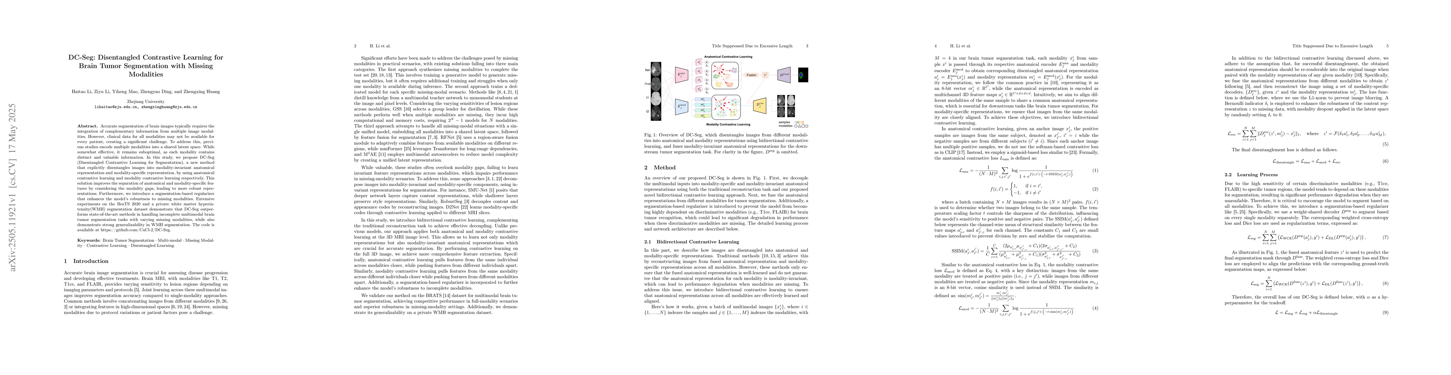
Accurate segmentation of brain images typically requires the integration of complementary information from multiple image modalities. However, clinical data for all modalities may not be available for every patient, creating a significant challenge. To address this, previous studies encode multiple modalities into a shared latent space. While somewhat effective, it remains suboptimal, as each modality contains distinct and valuable information. In this study, we propose DC-Seg (Disentangled Contrastive Learning for Segmentation), a new method that explicitly disentangles images into modality-invariant anatomical representation and modality-specific representation, by using anatomical contrastive learning and modality contrastive learning respectively. This solution improves the separation of anatomical and modality-specific features by considering the modality gaps, leading to more robust representations. Furthermore, we introduce a segmentation-based regularizer that enhances the model's robustness to missing modalities. Extensive experiments on the BraTS 2020 and a private white matter hyperintensity(WMH) segmentation dataset demonstrate that DC-Seg outperforms state-of-the-art methods in handling incomplete multimodal brain tumor segmentation tasks with varying missing modalities, while also demonstrate strong generalizability in WMH segmentation. The code is available at https://github.com/CuCl-2/DC-Seg.
AI Key Findings
Generated Jun 08, 2025
Methodology — What approach did the authors take?
The proposed method, DC-Seg, disentangles brain images into modality-invariant anatomical representation and modality-specific representation using anatomical contrastive learning and modality contrastive learning respectively. It also introduces a segmentation-based regularizer for handling missing modalities.
Key Results — What are the main findings?
- DC-Seg outperforms state-of-the-art methods in incomplete multimodal brain tumor segmentation tasks with varying missing modalities.
- The method demonstrates strong generalizability in white matter hyperintensity (WMH) segmentation.
Significance — Why does this research matter?
This research is important as it addresses the challenge of missing modalities in brain tumor segmentation, improving the accuracy and robustness of medical image analysis, which can lead to better clinical decision-making.
Technical Contribution — What is the technical contribution?
The disentangled contrastive learning approach for separating anatomical and modality-specific features, along with the segmentation-based regularizer for missing modality handling.
Novelty — What is new about this work?
Unlike previous methods that encode multiple modalities into a shared latent space, DC-Seg explicitly disentangles images into modality-invariant and modality-specific representations, leading to more robust segmentation performance.
Limitations — What are the limitations of this study?
- The paper does not discuss potential limitations or challenges encountered during the research.
- No information on scalability or computational resource requirements for the proposed method.
Future Work — What did the authors propose for future work?
- Explore the applicability of DC-Seg to other medical image segmentation tasks.
- Investigate methods to further reduce dependency on complete multimodal data.
How to Cite This Paper
@article{li2025dc,
  title         = {DC-Seg: Disentangled Contrastive Learning for Brain Tumor Segmentation
  with Missing Modalities},
  author        = {Li, Haitao and Mao, Yiheng and Huang, Zhengxing and others},
  year          = {2025},
  eprint        = {2505.11921},
  archivePrefix = {arXiv},
  primaryClass  = {cs.CV},
}Li, H., Mao, Y., Huang, Z., Ding, Z., & Li, Z. (2025). DC-Seg: Disentangled Contrastive Learning for Brain Tumor Segmentation
with Missing Modalities. arXiv. https://arxiv.org/abs/2505.11921Li, Haitao, et al. "DC-Seg: Disentangled Contrastive Learning for Brain Tumor Segmentation
with Missing Modalities." arXiv, 2025, arxiv.org/abs/2505.11921.PDF Preview
Citation Network
Current paper (gray), citations (green), references (blue)
Display is limited for performance on very large graphs.
Similar Papers
Found 4 papersM3AE: Multimodal Representation Learning for Brain Tumor Segmentation with Missing Modalities
Dong Wei, Liansheng Wang, Hong Liu et al.
Latent Correlation Representation Learning for Brain Tumor Segmentation with Missing MRI Modalities
Analyzing Deep Learning Based Brain Tumor Segmentation with Missing MRI Modalities
Shen Wang, Benteng Ma, Yushi Wang
No citations found for this paper.
Comments (0)