Discriminating retinal microvascular and neuronal differences related to migraines: Deep Learning based Crossectional Study
Publication
Metrics
AI Quick Summary
This study uses deep learning models to analyze retinal images and differentiate between migraineurs and non-migraineurs, finding that convolutional neural networks effectively discriminate through microvascular differences but less so through neuronal differences. The best performance was noted with color fundus photography, while optical coherence tomography did not significantly enhance model accuracy.
Paper Preview
Abstract
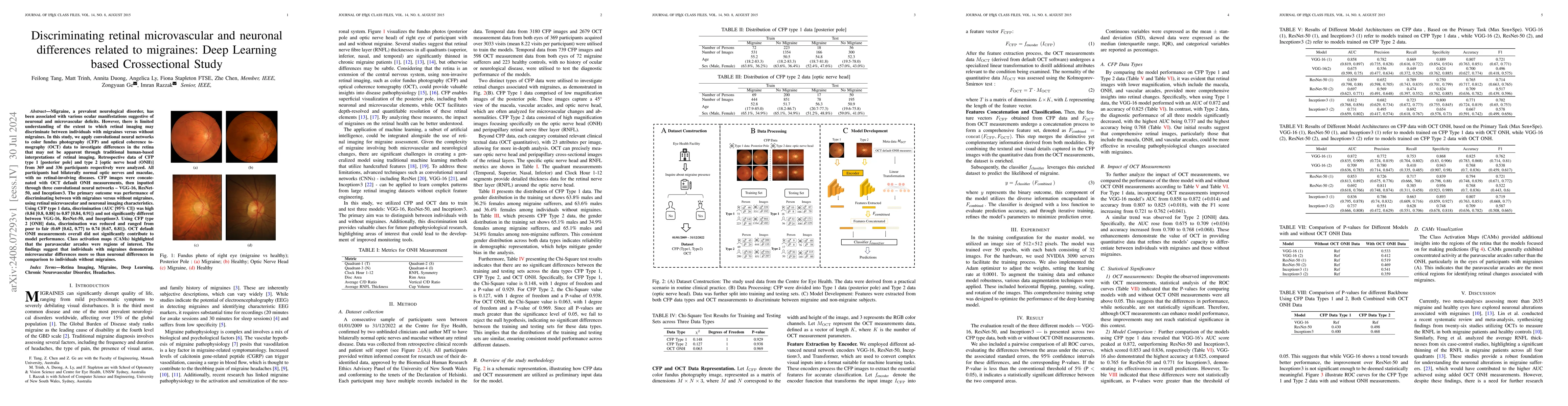
Migraine, a prevalent neurological disorder, has been associated with various ocular manifestations suggestive of neuronal and microvascular deficits. However, there is limited understanding of the extent to which retinal imaging may discriminate between individuals with migraines versus without migraines. In this study, we apply convolutional neural networks to color fundus photography (CFP) and optical coherence tomography (OCT) data to investigate differences in the retina that may not be apparent through traditional human-based interpretations of retinal imaging. Retrospective data of CFP type 1 [posterior pole] and type 2 [optic nerve head (ONH)] from 369 and 336 participants respectively were analyzed. All participants had bilaterally normal optic nerves and maculae, with no retinal-involving diseases. CFP images were concatenated with OCT default ONH measurements, then inputted through three convolutional neural networks - VGG-16, ResNet-50, and Inceptionv3. The primary outcome was performance of discriminating between with migraines versus without migraines, using retinal microvascular and neuronal imaging characteristics. Using CFP type 1 data, discrimination (AUC [95% CI]) was high (0.84 [0.8, 0.88] to 0.87 [0.84, 0.91]) and not significantly different between VGG-16, ResNet-50, and Inceptionv3. Using CFP type 2 [ONH] data, discrimination was reduced and ranged from poor to fair (0.69 [0.62, 0.77] to 0.74 [0.67, 0.81]). OCT default ONH measurements overall did not significantly contribute to model performance. Class activation maps (CAMs) highlighted that the paravascular arcades were regions of interest. The findings suggest that individuals with migraines demonstrate microvascular differences more so than neuronal differences in comparison to individuals without migraines.
AI Key Findings
Get AI-generated insights about this paper's methodology, results, significance, and more — seven facets brought into focus.
Impact
Authors
PDF Preview
Key Terms
Citation Network
Current paper (gray), citations (green), references (blue)
Display is limited for performance on very large graphs.
Discussion 0