The study presents a compact, dual-function optical device integrating fundus photography and refractive error detection using a coaxial optical design and deep learning algorithms.
Dual-Modality Computational Ophthalmic Imaging with Deep Learning and Coaxial Optical Design
Publication
Metrics
AI Quick Summary
This study introduces a compact dual-modality ophthalmic imaging device combining fundus photography and refractive error detection, utilizing a coaxial optical design and deep learning for precise pupil segmentation. The system achieves high-precision pupil localization and reliable refractive estimation, presenting a scalable solution for efficient eye screening in community health settings.
Quick Answers
What is "Dual-Modality Computational Ophthalmic Imaging with Deep Learning and Coaxial Optical Design" about?
This study introduces a compact dual-modality ophthalmic imaging device combining fundus photography and refractive error detection, utilizing a coaxial optical design and deep learning for precise pupil segmentation. The system achieves high-precision pupil localization and reliable refractive estimation, presenting a scalable solution for efficient eye screening in community health settings.
What methodology did the authors use?
The study presents a compact, dual-function optical device integrating fundus photography and refractive error detection using a coaxial optical design and deep learning algorithms. More in Methodology →
What are the key results?
High-precision pupil localization (EDE = 2.8 px, mIoU = 0.931) achieved with Dense-U-Net-based algorithm. — Reliable refractive estimation with a mean absolute error below 5%. More in Key Results →
Why is this work significant?
This research offers a promising solution for rapid, intelligent, and scalable ophthalmic screening, particularly suitable for community health settings, addressing the growing burden of myopia and retinal diseases. More in Significance →
What are the main limitations?
Limited by commercial lens components, unable to fully correct high-order aberrations. — Additional mirrors required due to compact assembly, compromising coaxial alignment and introducing edge-related dispersion. More in Limitations →
Abstract
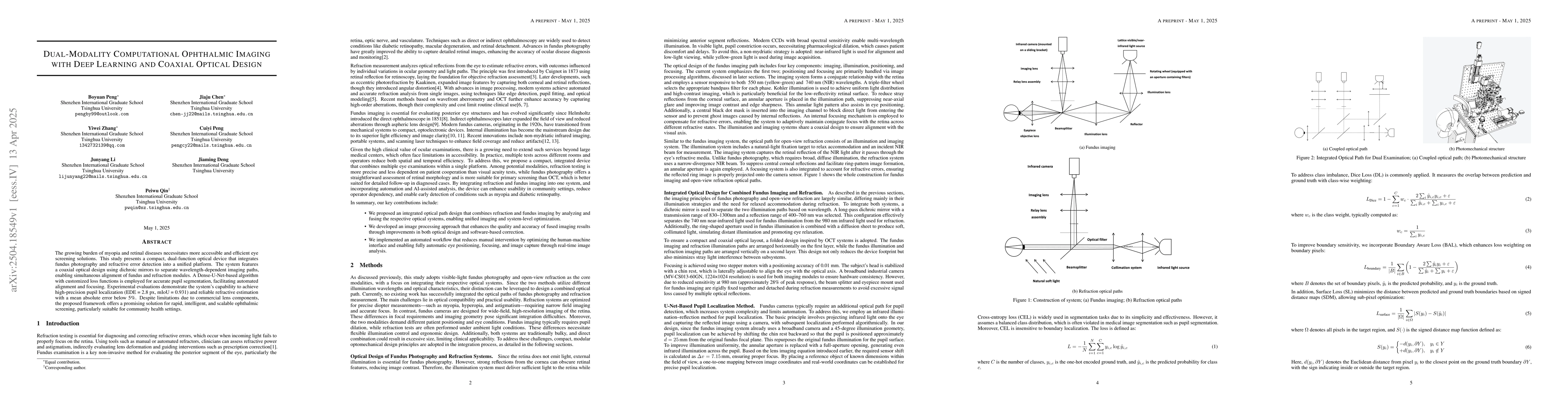
The growing burden of myopia and retinal diseases necessitates more accessible and efficient eye screening solutions. This study presents a compact, dual-function optical device that integrates fundus photography and refractive error detection into a unified platform. The system features a coaxial optical design using dichroic mirrors to separate wavelength-dependent imaging paths, enabling simultaneous alignment of fundus and refraction modules. A Dense-U-Net-based algorithm with customized loss functions is employed for accurate pupil segmentation, facilitating automated alignment and focusing. Experimental evaluations demonstrate the system's capability to achieve high-precision pupil localization (EDE = 2.8 px, mIoU = 0.931) and reliable refractive estimation with a mean absolute error below 5%. Despite limitations due to commercial lens components, the proposed framework offers a promising solution for rapid, intelligent, and scalable ophthalmic screening, particularly suitable for community health settings.
Key Findings, in focus
Seven facets of this paper, analysed and brought into focus by AI.
This research offers a promising solution for rapid, intelligent, and scalable ophthalmic screening, particularly suitable for community health settings, addressing the growing burden of myopia and retinal diseases.
- High-precision pupil localization (EDE = 2.8 px, mIoU = 0.931) achieved with Dense-U-Net-based algorithm.
- Reliable refractive estimation with a mean absolute error below 5%.
- Lightweight model with a parameter size of only 0.25 million, suitable for real-time processing on industrial devices.
This research offers a promising solution for rapid, intelligent, and scalable ophthalmic screening, particularly suitable for community health settings, addressing the growing burden of myopia and retinal diseases.
The introduction of a Dense-U-Net-based segmentation network for accurate ocular surface structure segmentation, combined with edge detection and unidirectional connected component extraction for rapid pupil boundary and center localization.
The proposed framework demonstrates improved segmentation performance through a customized training strategy with a combination of loss functions sensitive to different pixel types, without major structural innovations in the network architecture.
- Limited by commercial lens components, unable to fully correct high-order aberrations.
- Additional mirrors required due to compact assembly, compromising coaxial alignment and introducing edge-related dispersion.
- Further optimization of optical and mechanical hardware, including custom lens design and aspheric lenses.
- Expansion of clinical validation through large-scale studies in real-world healthcare environments.
Impact
Authors
PDF Preview
Citation Network
Current paper (gray), citations (green), references (blue)
Display is limited for performance on very large graphs.
Discussion 0