Trained a single UNet architecture at five downsampling ratios per dataset across five fundus datasets, keeping all other hyperparameters fixed, and evaluated performance using Dice and a width‑stratified sensitivity metric that separates thin, medium, and thick vessel detection.
Effect of Input Resolution on Retinal Vessel Segmentation Performance: An Empirical Study Across Five Datasets
Publication
Metrics
Quick Answers
What methodology did the authors use?
Trained a single UNet architecture at five downsampling ratios per dataset across five fundus datasets, keeping all other hyperparameters fixed, and evaluated performance using Dice and a width‑stratified sensitivity metric that separates thin, medium, and thick vessel detection. More in Methodology →
What are the key results?
For high‑resolution datasets (HRF, FIVES), downsampling consistently improves thin vessel sensitivity by up to ~9.4 percentage points while Dice remains stable. — For low‑to‑mid resolution datasets (DRIVE, STARE, CHASE_DB1), native resolution yields the best thin vessel sensitivity; any downsampling degrades it by up to 15.8 percentage points. More in Key Results →
Why is this work significant?
Demonstrates that input resolution choice must be tailored to native image size and encoder capacity, and that width‑stratified metrics are essential for fair cross‑dataset comparison, informing future retinal vessel segmentation benchmarks and clinical deployment. More in Significance →
What are the main limitations?
Only a single four‑stage UNet architecture was evaluated, limiting generality to other network designs. — Five‑fold cross‑validation with only five folds reduces statistical power and may not match official train/test splits. More in Limitations →
Abstract
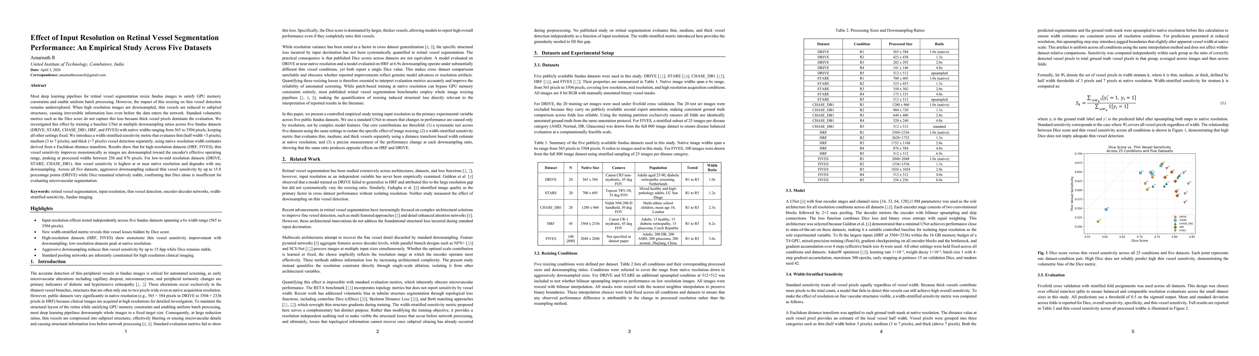
Most deep learning pipelines for retinal vessel segmentation resize fundus images to satisfy GPU memory constraints and enable uniform batch processing. However, the impact of this resizing on thin vessel detection remains underexplored. When high resolution images are downsampled, thin vessels are reduced to subpixel structures, causing irreversible information loss even before the data enters the network. Standard volumetric metrics such as the Dice score do not capture this loss because thick vessel pixels dominate the evaluation. We investigated this effect by training a baseline UNet at multiple downsampling ratios across five fundus datasets (DRIVE, STARE, CHASE_DB1, HRF, and FIVES) with native widths ranging from 565 to 3504 pixels, keeping all other settings fixed. We introduce a width-stratified sensitivity metric that evaluates thin (half-width <3 pixels), medium (3 to 7 pixels), and thick (>7 pixels) vessel detection separately, using native resolution width estimates derived from a Euclidean distance transform. Results show that for high-resolution datasets (HRF, FIVES), thin vessel sensitivity improves monotonically as images are downsampled toward the encoder's effective operating range, peaking at processed widths between 256 and 876 pixels. For low-to-mid resolution datasets (DRIVE, STARE, CHASE_DB1), thin vessel sensitivity is highest at or near native resolution and degrades with any downsampling. Across all five datasets, aggressive downsampling reduced thin vessel sensitivity by up to 15.8 percentage points (DRIVE) while Dice remained relatively stable, confirming that Dice alone is insufficient for evaluating microvascular segmentation.
Key Findings, in focus
Seven facets of this paper, analysed and brought into focus by AI.
Demonstrates that input resolution choice must be tailored to native image size and encoder capacity, and that width‑stratified metrics are essential for fair cross‑dataset comparison, informing future retinal vessel segmentation benchmarks and clinical deployment.
- For high‑resolution datasets (HRF, FIVES), downsampling consistently improves thin vessel sensitivity by up to ~9.4 percentage points while Dice remains stable.
- For low‑to‑mid resolution datasets (DRIVE, STARE, CHASE_DB1), native resolution yields the best thin vessel sensitivity; any downsampling degrades it by up to 15.8 percentage points.
- Dice score alone does not capture these resolution effects, as high Dice can coexist with poor thin vessel detection.
Demonstrates that input resolution choice must be tailored to native image size and encoder capacity, and that width‑stratified metrics are essential for fair cross‑dataset comparison, informing future retinal vessel segmentation benchmarks and clinical deployment.
Introduction of a width‑stratified sensitivity metric that isolates thin vessel detection performance and a systematic empirical framework linking input resolution to encoder receptive field across multiple datasets.
First controlled investigation showing that downsampling can improve microvascular detection on high‑resolution fundus images, revealing a resolution‑dependent trade‑off that contradicts common practice of uniform 512×512 resizing, and providing a new metric for cross‑dataset comparability.
- Only a single four‑stage UNet architecture was evaluated, limiting generality to other network designs.
- Five‑fold cross‑validation with only five folds reduces statistical power and may not match official train/test splits.
- Extend the study to architectures with larger receptive fields, dilated convolutions, vision transformers, or full‑resolution pathways to assess whether the effective operating range shifts.
- Combine width‑stratified sensitivity with topology‑aware metrics (e.g., clDice) and evaluate on full FIVES dataset and other disease‑specific datasets.
Discussion 0