Estimation of mitral valve hinge point coordinates -- deep neural net for echocardiogram segmentation
Publication
Metrics
AI Quick Summary
This paper proposes a fully automatic deep learning method using a U-Net to segment cardiac chambers in echocardiograms and extract mitral valve hinge points, achieving median absolute errors of 1.35 mm and 0.75 mm for x- and y-coordinates respectively. The method demonstrates improved accuracy over traditional feature-based detection.
Paper Preview
Abstract
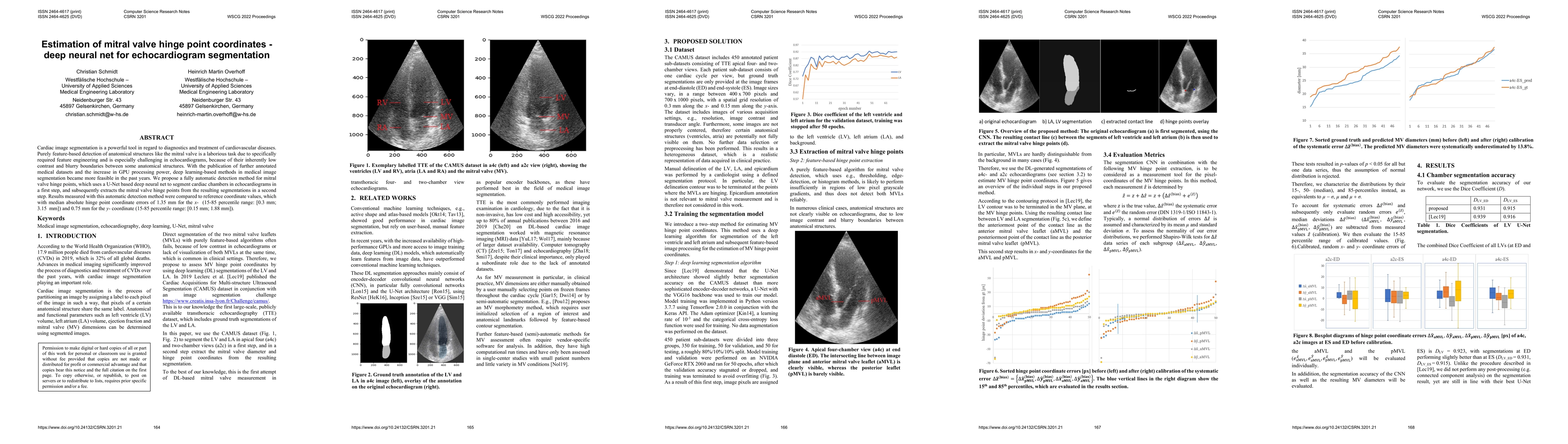
Cardiac image segmentation is a powerful tool in regard to diagnostics and treatment of cardiovascular diseases. Purely feature-based detection of anatomical structures like the mitral valve is a laborious task due to specifically required feature engineering and is especially challenging in echocardiograms, because of their inherently low contrast and blurry boundaries between some anatomical structures. With the publication of further annotated medical datasets and the increase in GPU processing power, deep learning-based methods in medical image segmentation became more feasible in the past years. We propose a fully automatic detection method for mitral valve hinge points, which uses a U-Net based deep neural net to segment cardiac chambers in echocardiograms in a first step, and subsequently extracts the mitral valve hinge points from the resulting segmentations in a second step. Results measured with this automatic detection method were compared to reference coordinate values, which with median absolute hinge point coordinate errors of 1.35 mm for the x- (15-85 percentile range: [0.3 mm; 3.15 mm]) and 0.75 mm for the y- coordinate (15-85 percentile range: [0.15 mm; 1.88 mm]).
AI Key Findings
Get AI-generated insights about this paper's methodology, results, significance, and more — seven facets brought into focus.
Impact
Paper Details
Authors
PDF Preview
Key Terms
Citation Network
Current paper (gray), citations (green), references (blue)
Display is limited for performance on very large graphs.
Discussion 0