ArxivLens
ArXiv physics.med-ph cond-mat.other

Event-sparse stack denoising for 4D-STEM applications

Espen Drath Bøjesen, Gregory Nordahl, Rebekka Klemmt

2512.10555 Published Dec 11, 2025
View PDF

Publication

Published: Dec 11, 2025
Categories: physics.med-ph, cond-mat.other

Metrics

Source: ArXiv

Quick Answers

What methodology did the authors use?

The research employs a combination of advanced electron microscopy techniques, specifically 4D-STEM, alongside machine learning algorithms for data analysis. It utilizes direct electron detectors with high temporal resolution to capture dynamic processes and applies principal component analysis (PCA) and DBSCAN clustering for pattern recognition and noise reduction. More in Methodology →

What are the key results?

Development of an event-driven 4D-STEM acquisition method with microsecond dwell times — Demonstration of sub-nanometer resolution for grain mapping in polycrystalline materials More in Key Results →

Why is this work significant?

This work advances the capabilities of electron microscopy for materials characterization by enabling high-resolution, low-dose imaging and precise strain analysis. It provides new tools for studying nanoscale phenomena in materials science. More in Significance →

What are the main limitations?

The method requires specialized equipment with high temporal resolution — Analysis complexity increases with larger datasets More in Limitations →

Paper Preview

Abstract

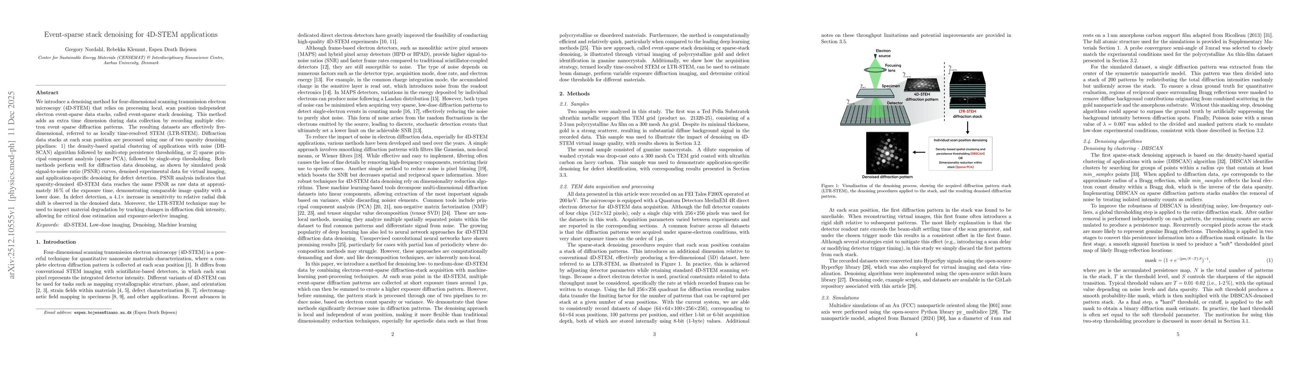
We introduce a denoising method for four-dimensional scanning transmission electron microscopy (4D-STEM) that relies on processing local, scan position-independent electron event-sparse data stacks, called event-sparse stack denoising. This method adds an extra time dimension during data collection by recording multiple electron event-sparse diffraction patterns. The resulting datasets are effectively five-dimensional, referred to as locally time-resolved STEM (LTR-STEM). Diffraction data stacks at each scan position are processed using one of two sparsity denoising pipelines: 1) the density-based spatial clustering of applications with noise (DBSCAN) algorithm followed by multi-step persistence thresholding, or 2) sparse principal component analysis (sparse PCA), followed by single-step thresholding. Both methods perform well for diffraction data denoising, as shown by simulated peak signal-to-noise ratio (PSNR) curves, denoised experimental data for virtual imaging, and application-specific denoising for defect detection. PSNR analysis indicates that sparsity-denoised 4D-STEM data reaches the same PSNR as raw data at approximately 16% of the exposure time, demonstrating comparable image quality with a lower dose. In defect detection, a 4.1x increase in sensitivity to relative radial disk shift is observed in the denoised data. Moreover, the LTR-STEM technique may be used to inspect material degradation by tracking changes in diffraction disk intensity, allowing for critical dose estimation and exposure-selective imaging.

Key Findings, in focus

Seven facets of this paper, analysed and brought into focus by AI.

AI-generated · verify against source Generated Dec 12, 2025
In focus · one-line synthesis

This work advances the capabilities of electron microscopy for materials characterization by enabling high-resolution, low-dose imaging and precise strain analysis.

01
MethodologyHow they did it

The research employs a combination of advanced electron microscopy techniques, specifically 4D-STEM, alongside machine learning algorithms for data analysis. It utilizes direct electron detectors with high temporal resolution to capture dynamic processes and applies principal component analysis (PCA) and DBSCAN clustering for pattern recognition and noise reduction.

02
ResultsWhat they found
  • Development of an event-driven 4D-STEM acquisition method with microsecond dwell times
  • Demonstration of sub-nanometer resolution for grain mapping in polycrystalline materials
  • Establishment of a reference-area-free strain mapping technique using precession electron diffraction
03
SignificanceWhy it matters

This work advances the capabilities of electron microscopy for materials characterization by enabling high-resolution, low-dose imaging and precise strain analysis. It provides new tools for studying nanoscale phenomena in materials science.

04
Technical ContributionsWhat's new technically

Creation of a sparse stack denoising framework for 4D-STEM data combined with machine learning approaches for enhanced pattern recognition and noise reduction.

05
NoveltyWhat sets it apart

The integration of event-driven acquisition with machine learning-based denoising represents a novel approach in 4D-STEM, enabling faster, more accurate analysis of complex materials datasets.

06
LimitationsWhat to be cautious about
  • The method requires specialized equipment with high temporal resolution
  • Analysis complexity increases with larger datasets
  • Limited to materials with sufficient electron transparency
07
Future WorkWhere it goes next
  • Integration with in-situ environmental cells for dynamic studies
  • Development of automated analysis pipelines
  • Exploration of 4D-STEM for biological samples

Paper Details

Paper ID: 2512.10555
Categories:
physics.med-ph cond-mat.other

How to Cite This Paper

Reference manager: RIS EndNote

Related Papers

4 found


Discussion 0

This website uses cookies to ensure you get the best experience. Learn more