The study repurposes a scanning Fabry-Perot interferometer (FPI) in spectral filtering mode for full-field Brillouin microscopy, combining it with light-sheet illumination for low-dose excitation and spatially resolved spectral acquisition.
Full-Field Brillouin Microscopy with a Scanning Fabry-Perot Interferometer
Publication
Metrics
Quick Answers
What methodology did the authors use?
The study repurposes a scanning Fabry-Perot interferometer (FPI) in spectral filtering mode for full-field Brillouin microscopy, combining it with light-sheet illumination for low-dose excitation and spatially resolved spectral acquisition. More in Methodology →
What are the key results?
Achieved millisecond-scale single-pixel dwell times and micrometer-scale spatial resolution with full 2D images acquired within one minute. — Demonstrated Brillouin emission imaging at selected frequency shifts, a capability unique to this system among spontaneous Brillouin techniques. More in Key Results →
Why is this work significant?
This research advances Brillouin microscopy by enabling high-speed, high-contrast full-field imaging, which could revolutionize mechanobiological studies and material characterization with non-contact, label-free techniques. More in Significance →
What are the main limitations?
Restricted field of view due to physical pinhole size and angular acceptance limitations of the FPI. — Spectral and angular distortions introduce uncertainties in viscosity measurements, requiring advanced correction algorithms. More in Limitations →
Abstract
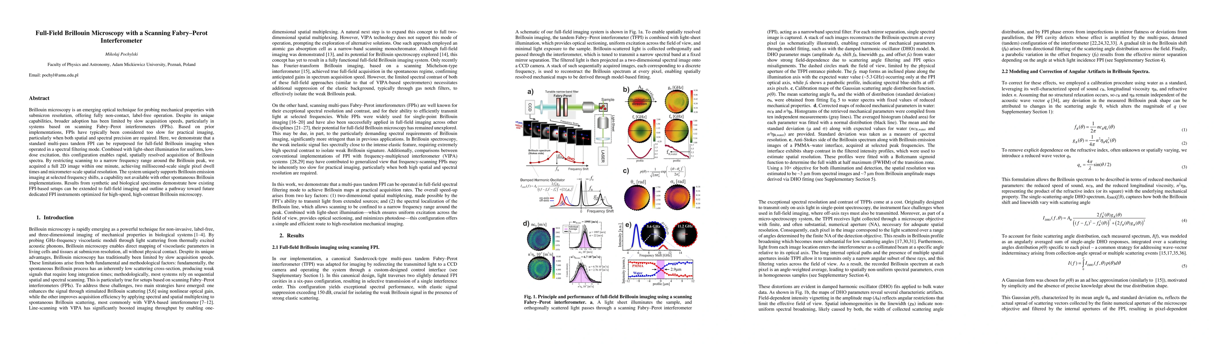
Brillouin microscopy is an emerging optical technique for probing mechanical properties with submicron resolution, offering fully non-contact, label-free operation. Despite its unique capabilities, broader adoption has been limited by slow acquisition speeds, particularly in systems based on scanning Fabry-Perot interferometers (FPIs). Based on prior implementations, FPIs have typically been considered too slow for practical imaging, particularly when both spatial and spectral precision are required. Here, we demonstrate that a standard multi-pass tandem FPI can be repurposed for full-field Brillouin imaging when operated in a spectral filtering mode. Combined with light-sheet illumination for uniform, low-dose excitation, this configuration enables rapid, spatially resolved acquisition of Brillouin spectra. By restricting scanning to a narrow frequency range around the Brillouin peak, we acquired a full 2D image within one minute, achieving millisecond-scale single pixel dwell times and micrometer-scale spatial resolution. The system uniquely supports Brillouin emission imaging at selected frequency shifts, a capability not available with other spontaneous Brillouin implementations. Results from synthetic and biological specimens demonstrate how existing FPI-based setups can be extended to full-field imaging and outline a pathway toward future dedicated FPI instruments optimized for high-speed, high-contrast Brillouin microscopy.
Key Findings, in focus
Seven facets of this paper, analysed and brought into focus by AI.
This research advances Brillouin microscopy by enabling high-speed, high-contrast full-field imaging, which could revolutionize mechanobiological studies and material characterization with non-contact, label-free techniques.
- Achieved millisecond-scale single-pixel dwell times and micrometer-scale spatial resolution with full 2D images acquired within one minute.
- Demonstrated Brillouin emission imaging at selected frequency shifts, a capability unique to this system among spontaneous Brillouin techniques.
- Enabled rapid mechanical property mapping of diverse samples including biological tissues and synthetic materials with high spectral contrast.
This research advances Brillouin microscopy by enabling high-speed, high-contrast full-field imaging, which could revolutionize mechanobiological studies and material characterization with non-contact, label-free techniques.
Integration of multi-pass tandem FPI with light-sheet illumination enables spatially resolved mechanical imaging without spatial scanning, achieving unprecedented acquisition speeds in Brillouin microscopy.
The first demonstration of full-field Brillouin imaging using a standard FPI in spectral filtering mode, combined with light-sheet illumination, overcoming prior limitations of slow acquisition speeds in FPI-based systems.
- Restricted field of view due to physical pinhole size and angular acceptance limitations of the FPI.
- Spectral and angular distortions introduce uncertainties in viscosity measurements, requiring advanced correction algorithms.
- Development of real-time interferometer performance monitoring systems for improved stability.
- Exploration of telecentric designs to enhance angular acceptance and field uniformity.
- Implementation of backscattering configurations for improved Brillouin linewidth measurements.
Discussion 0