Quick Answers
What methodology did the authors use?
The research employs a comprehensive analysis of spatial transcriptomics and histopathology data integration using deep learning models, including generative and cross-modal approaches to align histological images with gene expression profiles. More in Methodology →
What are the key results?
Development of foundation models that effectively integrate spatial transcriptomics with histopathology images — Establishment of cross-modal alignment techniques for accurate gene expression prediction from histology More in Key Results →
Why is this work significant?
This research advances the field by enabling more accurate prediction of gene expression from histopathological images, enhancing diagnostic capabilities and personalized treatment strategies in precision medicine. More in Significance →
What are the main limitations?
Current models may struggle with complex histological patterns and rare tissue types — Integration of multi-omics data remains challenging due to data heterogeneity More in Limitations →
Paper Preview
Abstract
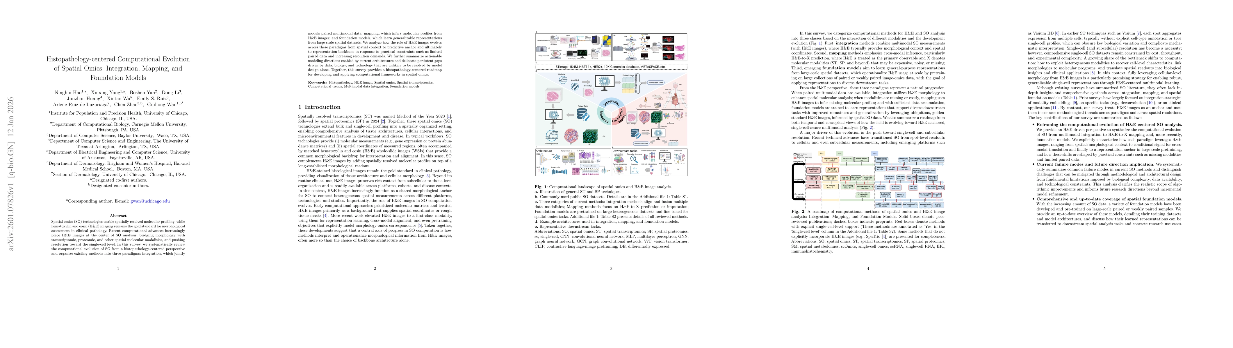
Spatial omics (SO) technologies enable spatially resolved molecular profiling, while hematoxylin and eosin (H&E) imaging remains the gold standard for morphological assessment in clinical pathology. Recent computational advances increasingly place H&E images at the center of SO analysis, bridging morphology with transcriptomic, proteomic, and other spatial molecular modalities, and pushing resolution toward the single-cell level. In this survey, we systematically review the computational evolution of SO from a histopathology-centered perspective and organize existing methods into three paradigms: integration, which jointly models paired multimodal data; mapping, which infers molecular profiles from H&E images; and foundation models, which learn generalizable representations from large-scale spatial datasets. We analyze how the role of H&E images evolves across these paradigms from spatial context to predictive anchor and ultimately to representation backbone in response to practical constraints such as limited paired data and increasing resolution demands. We further summarize actionable modeling directions enabled by current architectures and delineate persistent gaps driven by data, biology, and technology that are unlikely to be resolved by model design alone. Together, this survey provides a histopathology-centered roadmap for developing and applying computational frameworks in SO.
AI Key Findings
Generated Jan 13, 2026
Methodology — What approach did the authors take?
The research employs a comprehensive analysis of spatial transcriptomics and histopathology data integration using deep learning models, including generative and cross-modal approaches to align histological images with gene expression profiles.
Key Results — What are the main findings?
- Development of foundation models that effectively integrate spatial transcriptomics with histopathology images
- Establishment of cross-modal alignment techniques for accurate gene expression prediction from histology
- Creation of benchmark datasets for evaluating spatial transcriptomics prediction methods
Significance — Why does this research matter?
This research advances the field by enabling more accurate prediction of gene expression from histopathological images, enhancing diagnostic capabilities and personalized treatment strategies in precision medicine.
Technical Contribution — What is the technical contribution?
Introduction of novel deep learning frameworks, including foundation models and cross-modal denoising techniques, for integrating spatial transcriptomics data with histopathological images.
Novelty — What is new about this work?
The work introduces a unified approach combining spatial transcriptomics with histopathology through advanced deep learning models, offering new insights into tumor microenvironment analysis and improving diagnostic accuracy.
Limitations — What are the limitations of this study?
- Current models may struggle with complex histological patterns and rare tissue types
- Integration of multi-omics data remains challenging due to data heterogeneity
Future Work — What did the authors propose for future work?
- Exploring advanced architectures for better handling of histological variability
- Investigating federated learning approaches for data privacy in multi-institutional studies
- Developing more robust benchmarking frameworks for spatial transcriptomics analysis
Paper Details
How to Cite This Paper
@article{luzuriaga2026histopathology,
  title         = {Histopathology-centered Computational Evolution of Spatial Omics: Integration, Mapping, and Foundation Models},
  author        = {Luzuriaga, Arlene Ruiz de and Yan, Boshen and Zhao, Chen and others},
  year          = {2026},
  eprint        = {2601.07826},
  archivePrefix = {arXiv},
  primaryClass  = {q-bio.GN},
}Luzuriaga, A., Yan, B., Zhao, C., Li, D., Ruiz, E., Wan, G., Huang, J., Hao, N., Wu, X., & Yang, X. (2026). Histopathology-centered Computational Evolution of Spatial Omics: Integration, Mapping, and Foundation Models. arXiv. https://arxiv.org/abs/2601.07826Luzuriaga, Arlene Ruiz de, et al. "Histopathology-centered Computational Evolution of Spatial Omics: Integration, Mapping, and Foundation Models." arXiv, 2026, arxiv.org/abs/2601.07826.PDF Preview
Similar Papers
Found 4 papersHistopathology-centered Computational Evolution of Spatial Omics: Integration, Mapping, and Foundation Models.
Zhao, Chen, Li, Dong, Huang, Junzhou et al.
Computational methods for spatial multi-omics integration.
Zhang, Zilong, Cui, Feifei, Wei, Leyi et al.
A visual-omics foundation model to bridge histopathology with spatial transcriptomics.
Yang, Yu, Wang, Guangyu, Chen, Weiqing et al.
Multimodal Spatial Omics: From Data Acquisition to Computational Integration
Esra Busra Isik, Yusuf Hakan Usta, Haozhe Liu et al.
Comments (0)