01
MethodologyHow they did it
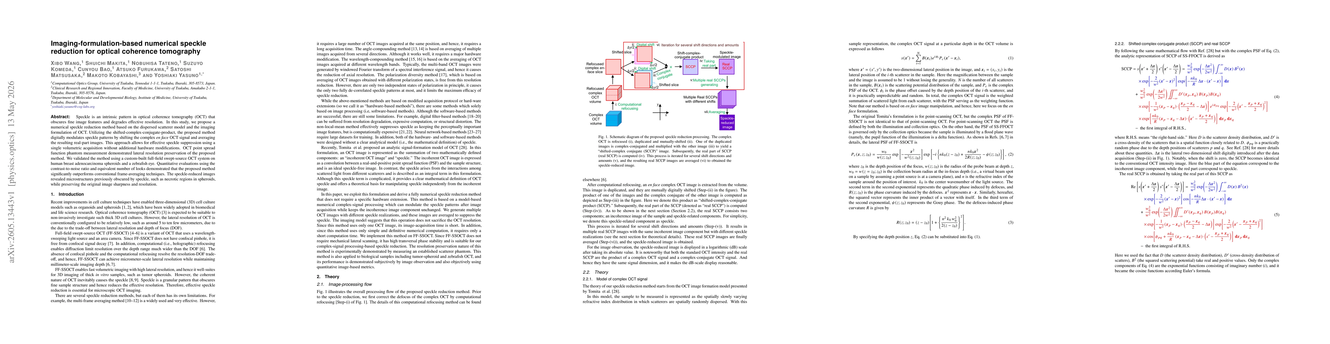
A model-based numerical speckle reduction for OCT leveraging the dispersed scatterer model and the imaging formulation of OCT. The approach uses the shifted-complex-conjugate-product to modulate speckle by shifting the complex en face OCT signal and averaging the resulting real-part images, after an initial computational refocusing step to correct defocus. It requires only a single volumetric acquisition and post-processing, applied to FF-SSOCT data, without hardware modifications, and validated against PSF phantoms and biological samples (tumor spheroids, zebrafish eye).
Discussion 0