01
MethodologyHow they did it
Brief description of the research methodology used
This paper reviews the historical development of electron microscopy in imaging ribosomes, emphasizing how cryo-EM has provided new insights into the assembly, biogenesis, and functional variations of ribosomes. It highlights the potential of cryogenic electron tomography to bridge cellular and structural biology.
This paper reviews the historical development of electron microscopy in imaging ribosomes, emphasizing how cryo-EM has provided new insights into the assembly, biogenesis, and functional variations of ribosomes. It highlights the potential of cryogenic electron tomography to bridge cellular and structural biology.
Brief description of the research methodology used More in Methodology →
Main finding 1 — Main finding 2 More in Key Results →
Why this research is important and its potential impact More in Significance →
Limitation 1 — Limitation 2 More in Limitations →
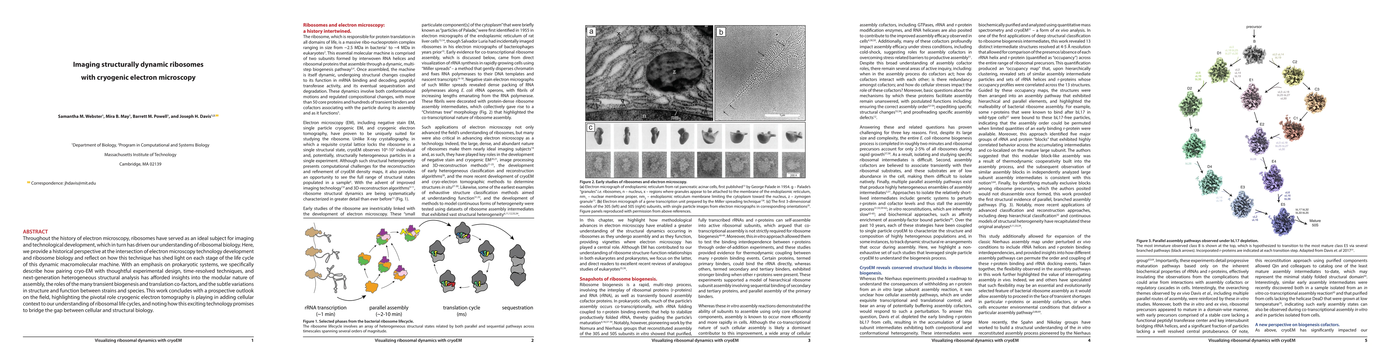
Throughout the history of electron microscopy, ribosomes have served as an ideal subject for imaging and technological development, which in turn has driven our understanding of ribosomal biology. Here, we provide a historical perspective at the intersection of electron microscopy technology development and ribosome biology and reflect on how this technique has shed light on each stage of the life cycle of this dynamic macromolecular machine. With an emphasis on prokaryotic systems, we specifically describe how pairing cryo-EM with clever experimental design, time-resolved techniques, and next-generation heterogeneous structural analysis has afforded insights into the modular nature of assembly, the roles of the many transient biogenesis and translation co-factors, and the subtle variations in structure and function between strains and species. The work concludes with a prospective outlook on the field, highlighting the pivotal role cryogenic electron tomography is playing in adding cellular context to our understanding of ribosomal life cycles, and noting how this exciting technology promises to bridge the gap between cellular and structural biology.
Seven facets of this paper, analysed and brought into focus by AI.
Why this research is important and its potential impact
Brief description of the research methodology used
Why this research is important and its potential impact
Main technical or theoretical contribution
What makes this work novel or different from existing research
Current paper (gray), citations (green), references (blue)
Display is limited for performance on very large graphs.
Discussion 0