01
MethodologyHow they did it
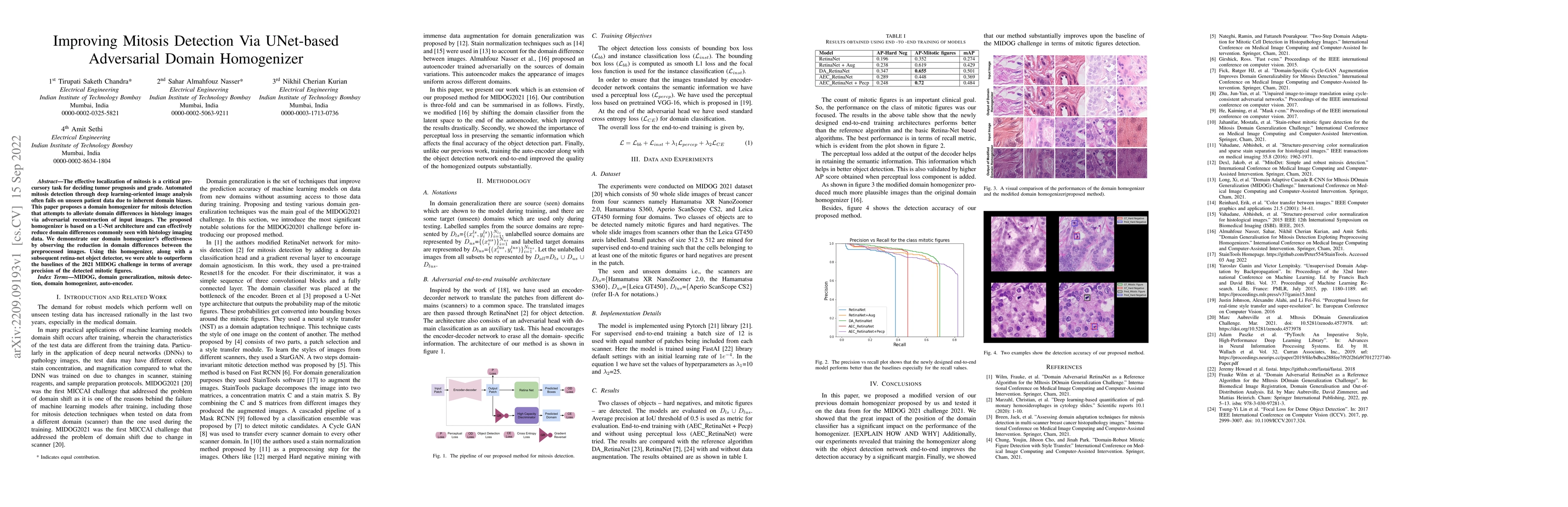
The research employs an encoder-decoder network for domain translation, followed by a RetinaNet for object detection. The architecture includes an adversarial head for domain classification, aiming to erase domain-specific information. The training involves bounding box loss, instance classification loss, perceptual loss for semantic information preservation, and standard cross-entropy loss for domain classification.
Discussion 0